3T MRI Scan in Surat: Advanced 3 Tesla MRI Imaging
High-Field MRI Technology for Detailed Diagnostic Imaging and Expert Radiology Evaluation
A 3T MRI Scan uses a 3 Tesla magnetic field to produce detailed images of internal body structures. MRI uses a powerful magnetic field, radio waves and computer technology rather than ionizing X-ray radiation to create diagnostic images. The higher field strength of a 3T system can provide strong image quality and may be particularly useful for examinations where detailed soft-tissue visualization is important.
At Super Scan Advanced Diagnostic & Radiology Centre, 3T MRI technology forms an important part of the centre’s advanced radiology setup in Surat. Super Scan states that its first 3T MRI system in Surat was introduced in 2014, reflecting its focus on bringing advanced radiology technology to the region.
What Is a 3T MRI Scan?
A 3T MRI, or 3 Tesla Magnetic Resonance Imaging scan, is an advanced form of MRI that operates with a magnetic field strength of 3 Tesla. It generates detailed cross-sectional images that allow radiologists to evaluate organs, soft tissues, joints, the brain, spine and other parts of the body.
MRI is a non-invasive imaging technique and does not use ionizing radiation. Depending on the clinical question, different MRI sequences may be selected to examine a particular body part or condition.
Why Is 3T MRI Used?
The main purpose of MRI is to provide detailed diagnostic information about structures that may be difficult to assess using other imaging methods. MRI can be particularly valuable for soft-tissue evaluation and is used for many neurological, musculoskeletal, abdominal, pelvic, vascular and other examinations.
A 3T system may offer advantages in situations where high-resolution imaging or specialized MRI sequences are required. However, the most appropriate MRI strength and protocol depend on the patient’s symptoms, clinical history and the examination requested by the treating doctor.
What Can a 3T MRI Scan Help Evaluate?
Depending on the doctor’s recommendation, MRI examinations can be performed for different parts of the body.
Brain and Neurological Imaging
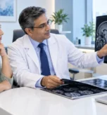
MRI can provide detailed images of the brain and surrounding structures. It is commonly used to evaluate various neurological concerns, including suspected tumors, stroke-related changes and other abnormalities. Specialized techniques may also be used when clinically appropriate.
Spine Imaging
MRI provides detailed views of the spinal cord, discs, nerves and surrounding soft tissues. It can help doctors evaluate conditions affecting the cervical, thoracic or lumbar spine.
Joint and Musculoskeletal Imaging
MRI can provide detailed images of muscles, ligaments, tendons, cartilage, joints and other soft-tissue structures. This makes MRI useful for evaluating many orthopedic and sports-related conditions.
Abdominal and Pelvic Imaging
MRI can be used to evaluate organs such as the liver, kidneys, pancreas, spleen and pelvic structures. Depending on the clinical requirement, specialized examinations may be performed for particular organs or conditions.
Blood Vessel Imaging
Specific MRI techniques such as MR angiography can be used to assess blood vessels. The exact technique depends on the body area and clinical indication.
What Are the Benefits of 3T MRI?
A major benefit of MRI is its ability to create detailed images without using ionizing radiation. MRI can provide strong soft-tissue contrast and can help physicians evaluate a wide range of conditions.
Potential benefits of a 3T MRI examination may include:
- Detailed imaging of soft tissues
- High-quality images for selected examinations
- Advanced MRI sequences for specific clinical questions
- Non-invasive diagnostic imaging
- No exposure to ionizing radiation
- Support for detailed evaluation of neurological and musculoskeletal structures
The actual diagnostic value depends on the body part being examined, MRI protocol, patient movement and the clinical question.
3T MRI vs Conventional MRI
The term 3T refers to the magnetic field strength of the MRI system. A higher field strength can provide a stronger signal that may be used to obtain detailed images or support specialized imaging protocols.
However, a higher Tesla value does not automatically mean that every patient or every examination requires 3T MRI. The appropriate scanner and protocol should be selected according to the patient’s clinical requirements and the radiologist’s or referring doctor’s recommendation.
Is 3T MRI Safe?
MRI does not use ionizing radiation, but the powerful magnetic field means that MRI safety screening is essential. Patients should inform the imaging team about implants, medical devices, metal fragments, previous surgeries and other relevant medical information before entering the MRI room.
Some implanted devices and metallic objects may be unsafe or require specific compatibility assessment. Patients should never assume that an implant is MRI-safe without confirmation from the appropriate medical or imaging team.
If contrast-enhanced MRI is planned, the radiology team may also ask about allergies and kidney-related medical history.
How Should You Prepare for a 3T MRI Scan?
Preparation depends on the type of MRI examination.
Before your appointment:
- Follow the specific instructions provided by the imaging centre.
- Bring previous relevant MRI, CT, X-ray or ultrasound reports and images when requested.
- Inform the radiology team about implants, pacemakers, surgical clips or other medical devices.
- Tell the team if you may be pregnant.
- Remove jewellery and other metallic objects before entering the MRI examination room.
- Wear comfortable clothing according to the centre’s instructions.
- Follow fasting instructions if contrast or sedation is required.
General MRI preparation can vary depending on the examination, so patients should follow the instructions provided for their specific appointment.
Super Scan’s existing 3T MRI guidance also advises patients to arrive before their appointment, avoid heavy cosmetics and ornaments, bring relevant previous imaging and medical records, and follow fasting instructions for contrast or sedation studies.
What Happens During the MRI Examination?
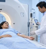
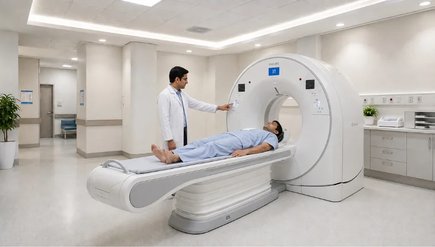
During an MRI examination, the patient lies on a movable examination table. The table gradually moves into the MRI scanner while the technologist acquires images using different sequences.
The scanner produces loud tapping or thumping sounds during imaging. Earplugs or headphones may be provided to reduce the noise. It is important to remain as still as possible because movement can affect image quality.
The technologist remains able to communicate with the patient during the examination. Some MRI studies may require an intravenous contrast agent when it is clinically indicated.
How Long Does a 3T MRI Take?
The duration of an MRI examination varies according to the body part being scanned, the number of sequences required and whether contrast is used. Some examinations may be relatively short, while more complex studies can take considerably longer.
Patients should therefore plan sufficient time for registration, preparation, screening and the examination itself.
Who Should Inform the MRI Team Before the Scan?
Patients should tell the radiology team before an MRI if they have:
- A pacemaker or implantable cardiac device
- Cochlear or other electronic implants
- Surgical clips or vascular devices
- Metal fragments in the body
- Previous major surgery
- Known kidney disease when contrast may be required
- Pregnancy or possible pregnancy
- Claustrophobia or significant anxiety
MRI compatibility can depend on the exact type and model of an implant or device, so appropriate screening and documentation are important.
Choosing a 3T MRI Centre in Surat
When selecting an MRI centre, patients should consider more than the scanner’s Tesla rating. Important factors include the experience of the radiology team, imaging protocols, patient safety procedures, reporting quality, appointment support and the centre’s ability to provide the examination recommended by the treating doctor.
Super Scan describes its Surat centres as providing advanced radiology technology with experienced radiologists and a comprehensive diagnostic imaging setup. Its current website lists centres at Majura Gate and Lal Darwaja in Surat.
3T MRI Scan at Super Scan, Surat
Super Scan Advanced Diagnostic & Radiology Centre has incorporated advanced MRI technology as part of its radiology services in Surat. The centre states that it introduced its first 3T MRI system in Surat in 2014 and has continued to focus on advanced diagnostic imaging technology and specialized radiology expertise.
For patients looking for 3T MRI in Surat, the appropriate examination should always be selected according to the doctor’s clinical recommendation and the patient’s individual diagnostic requirements.
Frequently Asked Questions About 3T MRI
Is 3T MRI better than a lower-field MRI?
A 3T MRI can provide strong signal and detailed imaging for many examinations, but the best MRI system depends on the specific clinical requirement. A higher Tesla rating is not automatically better for every examination.
Does 3T MRI use radiation?
No. MRI uses a magnetic field and radiofrequency energy rather than ionizing X-ray radiation.
Can I eat before a 3T MRI?
It depends on the type of examination. Some MRI studies require fasting, particularly when contrast or sedation is involved. Follow the instructions provided by the imaging centre.
Can I have an MRI with a metal implant?
Not every implant is automatically unsafe, but the exact device must be assessed for MRI compatibility. Always inform the MRI team before the examination.
Is contrast always required for MRI?
No. Contrast is used only when clinically indicated. The radiologist or referring doctor determines whether contrast is appropriate for the particular examination.
What should I bring for my MRI appointment?
Bring your prescription or referral, relevant previous imaging and reports, and any requested medical documents. Super Scan’s patient guidance specifically recommends bringing previous relevant X-ray, ultrasound, CT and MR studies and other case-related records.
Final Takeaway
A 3T MRI Scan is an advanced diagnostic imaging technique that can provide detailed views of many body structures without ionizing radiation. Its usefulness depends on the clinical indication, appropriate imaging protocol and expert interpretation. For patients in Surat, Gujarat and nearby regions, choosing an imaging centre with suitable MRI technology, experienced radiology professionals and appropriate patient-safety processes can help support a reliable diagnostic experience.
If your doctor has recommended a 3T MRI, discuss the specific examination, preparation requirements and any medical implants or conditions with the imaging centre before your appointment.
Medical Disclaimer: This article is intended for general educational information and does not replace advice from a qualified doctor or radiologist. The need for MRI, the appropriate protocol, contrast use and interpretation should be determined according to the individual patient’s clinical situation.