Understand the differences between mammography, breast ultrasound and breast MRI, and learn how age, symptoms, breast density and personal risk influence the choice of examination.
When it comes to breast imaging, many women wonder whether they need a mammogram, breast ultrasound or breast MRI. Although all three examinations help doctors evaluate breast health, each uses a different imaging technique and provides different information.
Mammography vs breast ultrasound vs MRI is not simply a question of which test is best. The appropriate examination depends on your age, symptoms, breast density, personal and family history, and whether the purpose is routine screening or investigating a specific concern.
Understanding these differences can help you have a more informed discussion with your doctor and choose the next step based on your individual clinical needs.
What Is Mammography?
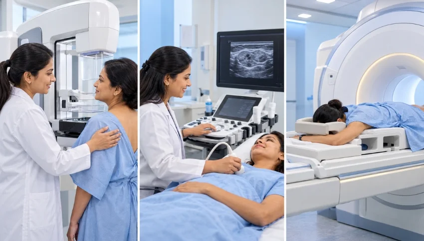
Mammography is a specialized X-ray examination that produces detailed images of breast tissue. It is an important method of breast cancer screening for women in the age and risk groups for whom screening is recommended. It is also used diagnostically when a symptom or previous imaging finding needs further evaluation.
During a mammogram, the breast is gently compressed between imaging plates for a few seconds while images are taken. Compression may cause temporary discomfort, but it helps spread the breast tissue and obtain useful images.
Mammography can reveal masses, changes in breast tissue and tiny calcium deposits called microcalcifications. Certain patterns of microcalcifications may be associated with early breast cancer and may need further assessment.
Does Mammography Use Radiation?
Yes. Mammography uses a low dose of X-ray radiation. When the examination is appropriately recommended, its expected benefits generally outweigh the small radiation-related risk.
The decision to undergo screening or diagnostic mammography should take into account age, symptoms, risk factors and the applicable clinical recommendations.
What Is Breast Ultrasound?
Breast ultrasound, also called breast sonography, uses high-frequency sound waves to create images of breast tissue. Unlike mammography, ultrasound does not use ionising radiation.
It is commonly used to evaluate a specific breast lump, a painful area or an abnormality found during a clinical examination or on another imaging test.
One important advantage of breast ultrasound is that it can help distinguish a fluid-filled cyst from a solid mass. It can also help radiologists examine a targeted area in greater detail.
Ultrasound is often particularly useful in younger women, whose breast tissue may be dense. However, it is not a universal replacement for mammography because some findings, including certain microcalcifications, are more readily detected on mammograms.
What Is Breast MRI?
Breast MRI uses a strong magnetic field, radio waves and a computer to produce detailed images of breast tissue. It does not use X-ray radiation.
Breast MRI is highly sensitive for detecting many breast abnormalities, but it is not routinely required for every woman. It is generally used for selected clinical situations, such as:
- Screening women who have a significantly elevated risk of breast cancer.
- Assessing the extent of a recently diagnosed breast cancer when additional information is needed.
- Evaluating selected findings that remain unclear after other imaging.
- Assessing breast implants or suspected implant complications in appropriate cases.
- Answering specific clinical questions when a radiologist or treating specialist considers MRI useful.
Many breast MRI examinations performed for cancer assessment use an intravenous contrast agent. The need for contrast, preparation requirements and suitability for MRI depend on the examination and the patient’s medical circumstances.
Mammography vs Breast Ultrasound vs MRI: Key Differences
The main difference is how each examination creates images and the clinical questions it is designed to answer.
Mammography
- Technology: X-ray imaging.
- Radiation: Uses a low dose of ionising radiation.
- Common role: Breast cancer screening and diagnostic assessment.
- Useful for: Masses, tissue changes and certain microcalcifications.
- Limitations: Dense breast tissue can make some abnormalities harder to see.
Breast Ultrasound
- Technology: Sound waves.
- Radiation: No ionising radiation.
- Common role: Evaluating a breast lump or targeted abnormality.
- Useful for: Characterising cysts and solid lesions and examining specific areas.
- Limitations: Does not replace mammography for all screening purposes and may not show certain abnormalities as clearly.
Breast MRI
- Technology: Magnetic field and radio waves.
- Radiation: No ionising radiation.
- Common role: Additional imaging for selected high-risk patients and specific diagnostic questions.
- Useful for: Detailed assessment of breast tissue and selected cases of known breast cancer.
- Limitations: May require contrast, can produce findings needing further investigation and is not necessary for every patient.
No single examination is best for every situation. The choice should be guided by the clinical question and the advice of the treating doctor or radiologist.
What Are Dense Breasts, and Do They Require Additional Imaging?
Breasts contain fatty tissue as well as fibrous and glandular tissue. When a relatively high proportion of fibrous and glandular tissue is present, the breasts are described as dense on a mammogram.
Dense breast tissue matters for two main reasons: it can make some abnormalities harder to detect on mammography, and it is associated with a higher risk of breast cancer compared with breasts that are not dense.
Depending on a woman’s overall risk, symptoms and mammography findings, a doctor may consider additional imaging such as ultrasound or MRI. However, dense breasts do not automatically mean that every woman needs a breast MRI. The potential benefits and limitations of supplemental screening should be discussed individually.
What If You Find a Lump in Your Breast?
Finding a breast lump can be worrying, but many breast lumps are benign. A new or persistent lump should nevertheless be evaluated rather than ignored.
The initial imaging test depends on factors such as age, examination findings, pregnancy or breastfeeding status, and the characteristics of the lump.
- Younger women: Targeted breast ultrasound is often an important first examination.
- Older women or selected clinical situations: Diagnostic mammography, sometimes combined with targeted ultrasound, may be recommended.
- Unclear or complex findings: Additional imaging or a biopsy may be advised, depending on the results.
The appropriate approach is not necessarily to undergo all available scans. A clinician should assess the concern and select the examinations that are most likely to provide useful information.
If a lump is accompanied by skin dimpling, new nipple inversion, bloody nipple discharge or other persistent breast changes, arrange prompt medical assessment.
What Does BI-RADS Mean on a Breast Imaging Report?
BI-RADS stands for Breast Imaging Reporting and Data System. It is a standardized system used by radiologists to describe breast imaging findings and communicate recommended next steps.
Depending on the category and the clinical context, recommendations may include routine screening, additional imaging, short-interval follow-up or a biopsy.
A BI-RADS category should not be interpreted in isolation. The full report, the actual imaging findings and the radiologist’s recommendation are important for understanding what should happen next.
If you receive a report that you do not understand, discuss it with your referring doctor or radiologist rather than drawing conclusions from the category number alone.
Which Breast Imaging Test Is Appropriate for Women With a Family History of Breast Cancer?
A strong family history of breast cancer, certain inherited genetic variants and other risk factors may affect when screening should begin and which examinations should be included.
For some women at elevated risk, a screening plan may include both mammography and breast MRI. Other women may need a different approach based on their age, estimated risk and medical history.
Not everyone with a relative who had breast cancer will require the same imaging schedule. Discuss your family history with a qualified healthcare professional, who can determine whether a formal risk assessment or genetic counselling is appropriate.
Can Breast Ultrasound Replace Mammography?
Not in every situation. Although ultrasound is useful for examining breast lumps and targeted abnormalities, it may not detect some findings that mammography can reveal, including certain microcalcifications.
For this reason, ultrasound is often used alongside mammography when additional information is needed. The tests are complementary rather than interchangeable.
Whether you need one or both examinations depends on the reason for imaging, your age, symptoms and risk profile.
Do You Always Need a Breast MRI After Mammography?
No. Breast MRI is an additional examination used when it is expected to answer a specific clinical question or provide useful information for screening or treatment planning.
It may be recommended for certain women at high risk, selected patients with a breast cancer diagnosis or situations where other imaging has not resolved a concern. It is not automatically required after a normal mammogram or simply because breast tissue is dense.
A doctor or radiologist can explain whether MRI is likely to add meaningful information in your particular situation.
Frequently Asked Questions (FAQs)
1. Which is better: mammography, ultrasound or breast MRI?
There is no single best test for every woman. Mammography is important for screening in appropriate age and risk groups, ultrasound is useful for evaluating many breast lumps, and MRI is valuable in selected high-risk and diagnostic situations.
2. Is breast ultrasound safer than mammography because it has no radiation?
Ultrasound does not use ionising radiation, but that does not make it a replacement for mammography in every situation. Each test has different strengths and limitations, and the appropriate examination depends on the clinical need.
3. Can a mammogram detect breast cancer early?
Mammography can detect certain breast cancers and suspicious changes before they cause noticeable symptoms. Screening recommendations depend on age, personal risk and applicable clinical guidance. No screening test detects every cancer.
4. Do dense breasts mean I need an MRI?
Not necessarily. Additional imaging may be considered depending on overall breast cancer risk, symptoms and mammography findings. Discuss the potential benefits and limitations of supplemental screening with your healthcare professional.
5. Does breast MRI use radiation?
No. Breast MRI uses a magnetic field and radio waves, not ionising radiation. Many breast MRI examinations use intravenous contrast, so the imaging team should review relevant medical information before the scan.
6. Which scan is usually done first for a breast lump?
The initial examination depends on age and clinical findings. Targeted ultrasound is often used first in younger women, while diagnostic mammography with or without ultrasound may be appropriate in other situations. A clinician should guide the choice.
7. Where can I enquire about mammography in Surat?
You can contact Super Scan at +91 95125 48000 or email info@superscan.in to enquire about mammography services and appointment availability.
Conclusion
Mammography, breast ultrasound and breast MRI each have a distinct role in breast imaging. Mammography is an important screening and diagnostic tool, ultrasound helps evaluate many targeted abnormalities, and MRI provides additional information in selected situations.
The right choice depends on your age, symptoms, breast density, personal and family history, and the reason for the examination. Rather than choosing a scan based on online comparisons alone, discuss your individual needs with your doctor or radiologist.
Also read,
- Diagnostic Imaging Services in Surat
- Colour Doppler
- Contrast Procedures
- 3D and 4D Sonography
- PET Scan
- Gamma Scan