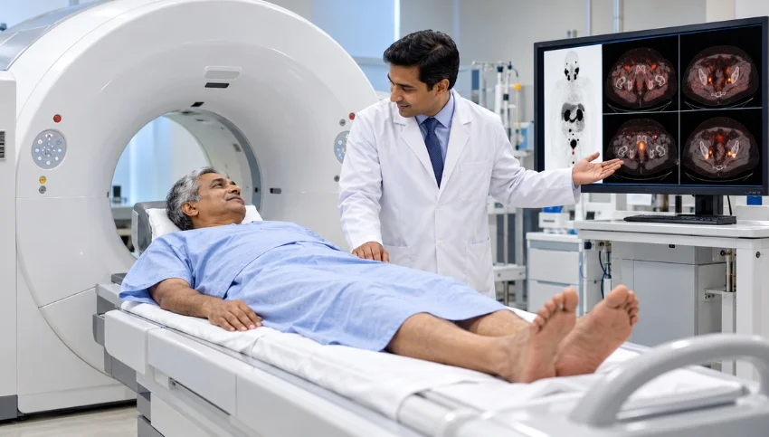
Learn when PSMA PET-CT may be recommended, what the scan can reveal, and how imaging findings help doctors plan prostate cancer care.
If you or a family member is being evaluated or treated for prostate cancer, your doctor may recommend a PSMA PET-CT scan to obtain more information about the location and extent of the disease. This specialized molecular imaging examination can help doctors assess prostate cancer in selected situations, including initial staging and suspected recurrence after treatment.
Unlike conventional imaging, PSMA PET-CT uses a targeted radioactive tracer that binds to prostate-specific membrane antigen (PSMA). The scan combines PET imaging with CT images to help identify areas of abnormal tracer uptake and assess their anatomical location.
For patients considering a PSMA PET-CT scan in Surat, understanding when the examination may be useful, what it can reveal, and how doctors interpret the results can make the imaging process easier to understand.
What Is a PSMA PET-CT Scan?
PSMA stands for Prostate-Specific Membrane Antigen, a protein found in high amounts on many prostate cancer cells. A PSMA PET-CT scan uses a radiotracer designed to target this protein.
After the tracer is administered, it travels through the body and may accumulate in areas where PSMA is expressed. The PET scanner detects the tracer distribution, while the CT component provides anatomical information to help locate the areas of uptake.
PSMA PET-CT is used for specific clinical questions related to prostate cancer. It is not a replacement for every other prostate examination, and the decision to perform it depends on the patient’s diagnosis, risk category, PSA level, previous treatment and the information needed by the treating team.
How Is PSMA PET-CT Different From an FDG PET Scan?
Both PSMA PET-CT and FDG PET-CT are forms of molecular imaging, but they use different tracers and assess different biological processes.
- PSMA PET-CT: Uses a tracer that targets PSMA and is particularly useful in appropriate prostate cancer assessments.
- FDG PET-CT: Uses fluorodeoxyglucose (FDG), a glucose-related tracer that helps evaluate metabolic activity in many types of disease.
PSMA PET-CT is often preferred for specific prostate cancer staging and recurrence questions because it targets a biological feature associated with many prostate cancer cells. However, not every prostate cancer lesion expresses PSMA equally, and no scan detects every tumour in every patient.
Your doctor will select the imaging examination according to the clinical question rather than assuming that one type of PET scan is suitable for all cancers.
When Is PSMA PET-CT Needed for Prostate Cancer?
A doctor may recommend PSMA PET-CT when the findings are expected to help clarify the extent of prostate cancer or influence treatment planning.
1. Initial Staging After a Prostate Cancer Diagnosis
After prostate cancer has been diagnosed, doctors assess whether it appears confined to the prostate or may have spread to other parts of the body.
For selected patients, particularly those with unfavourable intermediate-risk or high-risk disease, PSMA PET-CT may help evaluate possible spread to lymph nodes, bones or other sites. The need for this scan depends on the cancer’s risk category and the clinical guidelines being followed.
Staging information can help the treating team discuss suitable management options, which may include surgery, radiotherapy, systemic treatment or a combination of approaches.
2. Investigating a Rising PSA Level After Treatment
Prostate-specific antigen (PSA) is a blood marker used in the assessment and follow-up of prostate cancer. After treatment, a rising PSA level may raise concern about persistent or recurrent disease.
In appropriate patients, PSMA PET-CT can help locate areas suspicious for recurrence, including disease in the prostate bed, lymph nodes or distant sites. The usefulness of imaging depends partly on the PSA level, its rate of change, previous treatment and the likelihood that the result will influence the next clinical decision.
A negative scan does not always rule out recurrent cancer, particularly when disease is too small or does not show sufficient tracer uptake.
3. Assessing Suspected Spread to Other Parts of the Body
When prostate cancer may have spread beyond the prostate, PSMA PET-CT can help evaluate areas throughout the imaged body.
Potential sites assessed include lymph nodes, bones and selected other organs. The findings may help the treating team understand the distribution of disease and consider whether additional investigations or changes to the treatment plan are appropriate.
4. Evaluating Suitability for PSMA-Targeted Treatment
In selected patients with advanced prostate cancer, PSMA imaging may help determine whether the disease shows sufficient target expression for a PSMA-directed radioligand treatment.
This assessment is only one part of treatment eligibility. The oncology and nuclear medicine teams also consider the patient’s clinical condition, previous treatments, laboratory results, organ function and the relevant treatment criteria.
What Can a PSMA PET-CT Scan Show?
A PSMA PET-CT examination may provide information about:
- The prostate or prostate bed: Areas suspicious for disease in the prostate or the region where the prostate was previously located.
- Lymph nodes: Possible involvement of pelvic or other lymph nodes.
- Bones: Areas of abnormal uptake that may require assessment for possible bone metastases.
- Other sites: Selected areas outside the pelvis that may be relevant to staging or recurrence.
- PSMA expression: The distribution and intensity of tracer uptake, which may be considered when assessing eligibility for certain targeted treatments.
The final interpretation depends on the complete scan, the tracer used and the patient’s medical history. Some abnormalities require further assessment, comparison with earlier imaging or correlation with other tests.
Does Every Bright Spot on PSMA PET-CT Mean Cancer?
No. Increased PSMA tracer uptake does not automatically mean that an area contains prostate cancer.
Some normal tissues show physiological uptake, and certain non-cancerous conditions or other tumours can also demonstrate uptake. Conversely, some prostate cancer lesions may show limited or undetectable uptake.
A specialist interprets the PET and CT images together with the patient’s PSA level, pathology results, symptoms, treatment history and previous imaging. Depending on the findings, the doctor may recommend follow-up imaging, additional tests or clinical correlation.
The scan report should therefore be understood as part of the overall medical assessment, not as an isolated diagnosis.
How Should You Prepare for a PSMA PET-CT Scan?
Preparation instructions can vary depending on the tracer, imaging protocol and individual medical circumstances. Follow the instructions provided by your imaging centre rather than applying preparation rules from a different type of PET scan.
Before your appointment:
- Share your prostate cancer diagnosis, recent PSA results and relevant medical reports.
- Inform the team about previous surgery, radiotherapy, hormone therapy, chemotherapy or other cancer treatments.
- Bring earlier scan reports and images when available.
- Tell the team about your current medicines, allergies and relevant medical conditions.
- Ask whether you need to follow any specific instructions regarding food, fluids or medicines.
Fasting requirements for PSMA PET-CT may differ from those for FDG PET-CT. Do not stop prescribed medicines or change treatment unless your treating team instructs you to do so.
What Happens During a PSMA PET-CT Examination?
The examination usually involves the following steps:
- Patient registration and review: The team confirms the referral, medical history and relevant reports.
- Tracer administration: A small amount of the prescribed radioactive tracer is administered, usually through an injection into a vein.
- Waiting period: The tracer is given time to distribute through the body. The duration depends on the tracer and the imaging protocol.
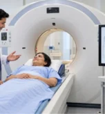
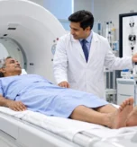
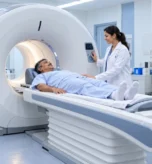
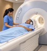
- Image acquisition: You lie on the scanner table while the PET and CT images are acquired. It is important to remain as still as possible.
- Image interpretation: A qualified specialist reviews the images and prepares a report for the referring doctor.
The scan itself is generally painless apart from the injection. The total appointment time varies according to the tracer, waiting period and scanning protocol. The imaging team will explain any additional instructions, including hydration and radiation-safety precautions after the examination.
Is PSMA PET-CT Safe?
PSMA PET-CT is a medical imaging examination that involves exposure to ionising radiation from the radiotracer and the CT component. The clinical team considers the expected diagnostic benefit, radiation exposure and individual circumstances when recommending the scan.
Tell your doctor and the imaging team if you are pregnant or think you may be pregnant, and provide information about relevant medical conditions and medicines. Follow the centre’s instructions regarding hydration, toileting and contact with others after the scan, where applicable.
Your imaging team can explain the precautions relevant to the specific tracer and protocol being used.
What Happens After the PSMA PET-CT Results?
The imaging specialist prepares a report describing the distribution of tracer uptake and any findings that may be relevant to the clinical question.
Your treating doctor interprets the report alongside the PSA trend, pathology, symptoms, previous treatment and other investigations. Depending on the results, the next step may involve continued monitoring, further tests, local treatment, systemic treatment or a multidisciplinary discussion.
A positive scan does not independently determine the treatment plan, and a negative scan does not exclude every possibility of disease. Treatment decisions should be made with the appropriate clinical team.
What Is PSMA Theranostics? Understanding Lu-177 Treatment
PSMA is important not only for imaging but also for certain targeted treatments. In selected patients, a PSMA-targeted radioactive medicine can deliver radiation to cells that express the target. This approach is known as PSMA-targeted radioligand therapy, a form of theranostics.
One example is Lutetium-177 (Lu-177) PSMA therapy, which may be considered for eligible patients with advanced prostate cancer. Before treatment, the specialist team assesses PSMA imaging and other clinical factors to determine whether the therapy is appropriate.
PSMA PET-CT and Lu-177 therapy serve different purposes: imaging helps assess where target-expressing disease may be present, while treatment delivers radiation to selected target-expressing cells. Not every patient with prostate cancer is eligible for Lu-177 therapy, and the decision requires specialist assessment.
Frequently Asked Questions
1. What is a PSMA PET-CT scan used for?
PSMA PET-CT is used in appropriate patients to help stage prostate cancer, investigate suspected recurrence and assess the distribution of PSMA-expressing disease. It may also help evaluate suitability for selected PSMA-targeted treatments.
2. Is PSMA PET-CT better than a regular FDG PET scan for prostate cancer?
PSMA PET-CT is specifically useful for many prostate cancer imaging questions because its tracer targets PSMA. FDG PET-CT evaluates glucose-related metabolic activity and may be more appropriate for other clinical questions. The choice depends on the cancer type and the purpose of imaging.
3. Can PSMA PET-CT detect recurrent prostate cancer when PSA rises?
It can help locate suspected recurrent disease in suitable patients. The chance of detecting disease depends on factors including the PSA level, PSA trend, tumour characteristics and previous treatment. A negative result does not completely rule out recurrence.
4. Does PSMA PET-CT show cancer in bones and lymph nodes?
It can identify areas suspicious for prostate cancer involvement in bones and lymph nodes when those areas demonstrate relevant tracer uptake. Findings must be interpreted by a specialist and correlated with other clinical information.
5. Do I need to fast before PSMA PET-CT?
Fasting requirements vary by tracer and imaging protocol. Do not assume that instructions for an FDG PET-CT apply to PSMA PET-CT. Follow the specific instructions provided by your imaging centre.
6. Is PSMA PET-CT used before Lu-177 PSMA therapy?
PSMA imaging is an important part of evaluating patients for PSMA-targeted radioligand therapy. However, treatment eligibility also depends on clinical assessment, previous treatment, laboratory results and other criteria.
7. Where can I enquire about a PSMA PET-CT scan in Surat?
You can contact Super Scan to ask about PET-CT services at its Khatodara centre and confirm whether the specific PSMA tracer and protocol required for your referral are available.
Conclusion
PSMA PET-CT has an important role in selected prostate cancer assessments, particularly initial staging, suspected recurrence and evaluation for certain targeted treatments. By combining molecular imaging with CT anatomy, it can provide information that helps the treating team understand the distribution of disease and plan the next steps.
If your doctor has recommended a PSMA PET-CT scan, discuss its purpose, expected benefits, limitations and preparation requirements with your treating team. For patients in Surat and South Gujarat, Super Scan can be contacted to confirm the availability of the required PET-CT examination.
Also read,
- Diagnostic Imaging Services in Surat
- Colour Doppler
- Contrast Procedures
- 3D and 4D Sonography
- PET Scan
- Gamma Scan