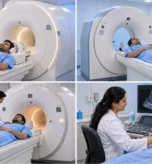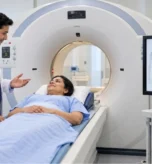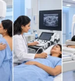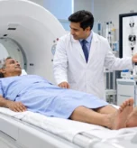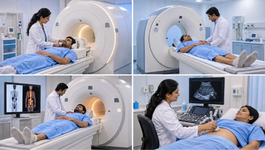
Understand the differences between MRI, CT, PET-CT and ultrasound, including their uses, limitations and radiation considerations, to learn why the right scan depends on your medical needs.
If your doctor has recommended an imaging test, you may wonder whether you need an MRI, CT scan, PET-CT or ultrasound. These examinations use different technologies and provide different types of information about the body. Understanding the differences between MRI vs CT vs PET-CT vs ultrasound can help you understand why your doctor has selected a particular investigation.
There is no single best scan for every medical condition. Ultrasound is useful for many abdominal organs and blood-flow assessments, CT provides fast and detailed images for many emergencies, MRI offers excellent soft-tissue detail, and PET-CT combines molecular information with anatomical imaging for selected conditions, particularly in cancer care. The appropriate test depends on your symptoms, medical history and the clinical question your doctor needs answered.
At Super Scan Imaging Centres, Surat, diagnostic imaging services include MRI, CT, ultrasound, Doppler and PET-CT, helping clinicians investigate a wide range of medical conditions.
What Is the Difference Between MRI, CT, PET-CT and Ultrasound?
The main difference is how each examination creates images and what information it can provide.
Ultrasound: Imaging With Sound Waves
Ultrasound, also called sonography, uses high-frequency sound waves to create images of internal structures. It does not use ionizing radiation and can provide real-time images during an examination.
Doctors commonly use ultrasound to assess the liver, gallbladder, kidneys, thyroid, uterus, ovaries and certain breast abnormalities. It is also widely used during pregnancy and to investigate selected lumps and swellings.
Doppler ultrasound provides additional information about blood flow through certain arteries and veins.
However, ultrasound has limitations. Air and bone can obstruct sound waves, and image quality can depend on the body area, bowel gas, body build and the examination technique. Some findings require further assessment with CT or MRI.
CT Scan: Fast, Detailed Cross-Sectional Imaging
A CT scan, or computed tomography scan, uses X-rays and computer processing to create detailed cross-sectional images of the body.
CT is particularly useful when doctors need images quickly. Depending on the clinical situation, it may help assess serious injuries, internal bleeding, lung conditions, kidney stones, acute abdominal problems and many bone abnormalities. Specialized CT examinations can also assess blood vessels and the coronary arteries.
Some CT examinations require contrast material to make certain structures easier to evaluate. The need for contrast depends on the reason for the scan.
CT uses ionizing radiation. Doctors consider the expected diagnostic benefit, the patient’s circumstances and appropriate dose-reduction techniques when deciding whether it is needed.
MRI Scan: Detailed Images of Soft Tissues
MRI, or magnetic resonance imaging, uses a strong magnetic field and radiofrequency signals to produce detailed images without ionizing radiation.
It is particularly valuable for assessing the brain, spinal cord, joints, ligaments, muscles and many abdominal, pelvic and prostate conditions. MRI may also be used in selected breast examinations and for characterising certain abnormalities found on other scans.
MRI is not automatically better than CT. It generally takes longer, and patients must be screened for implants, metal fragments and other potential magnetic-field safety concerns. Some examinations also require contrast material.
PET-CT: Information About Biological Activity and Anatomy
PET-CT combines positron emission tomography (PET) with computed tomography (CT). The PET component uses a radiotracer to show aspects of biological activity, while the CT component helps locate the areas of uptake within the body.
PET-CT is particularly useful in selected cancer-related situations, including assessing disease extent, evaluating response to treatment and investigating suspected recurrence. Its usefulness depends on the type of disease, the tracer used and the clinical question.
Different PET tracers target different biological processes. For example, FDG PET-CT evaluates glucose-related metabolic activity, PSMA PET-CT is used in appropriate prostate cancer assessments, and DOTA PET-CT can help evaluate selected neuroendocrine tumors.
PET-CT involves radiation from the radiotracer and the CT component. It is not routinely required for every patient with suspected or confirmed cancer.
MRI vs CT Scan: Which One Is More Suitable?
MRI and CT provide different information, so the right choice depends on the body part and suspected condition.
MRI may be preferred when:
- Detailed assessment of the brain, spinal cord or nerves is needed.
- A ligament, cartilage or other soft-tissue injury is suspected.
- Certain liver, pelvic or prostate abnormalities require further characterisation.
- A suitable examination without ionizing radiation is desirable and MRI is clinically appropriate.
CT may be preferred when:
- Rapid imaging is required in an emergency.
- Detailed assessment of the lungs, certain bone injuries or kidney stones is needed.
- Doctors need to evaluate particular chest or abdominal conditions.
- A CT angiography examination is appropriate to assess blood vessels.
Neither test is universally superior. In some situations, doctors may recommend both because each examination can answer a different clinical question.
MRI vs Ultrasound: What Is the Difference?
Ultrasound is often used as an initial examination because it is widely available, provides real-time images and does not use ionizing radiation.
MRI provides more detailed cross-sectional images for many soft-tissue and deep-organ questions, but it usually takes longer and may not be the first test required.
For example, ultrasound may be suitable for an initial assessment of certain abdominal symptoms. If the findings are uncertain or more detailed characterisation is needed, a doctor may recommend MRI or another investigation.
The choice depends on the suspected condition, the area being examined and whether the initial scan provides enough information.
CT Scan vs Ultrasound: When Is Each Used?
Both tests are useful, but they work differently.
Ultrasound is commonly used to examine the gallbladder, liver, kidneys, pelvic organs, thyroid and blood flow. It is also frequently used during pregnancy when clinically appropriate.
CT may be more suitable when doctors need a wider, detailed assessment of the chest or abdomen, rapid evaluation of trauma or investigation of conditions that ultrasound cannot adequately demonstrate.
CT uses ionizing radiation, whereas ultrasound does not. However, avoiding radiation is not the only consideration: the chosen examination must be capable of answering the medical question accurately.
PET-CT vs CT Scan: Why Are They Different?
A CT scan primarily provides anatomical information about the structures inside the body. PET-CT adds information about tracer uptake, which can reflect particular biological or metabolic processes.
For selected cancers, PET-CT may help doctors assess disease distribution or response to treatment. A conventional CT may be sufficient for other conditions or follow-up situations.
PET-CT is not automatically required whenever a CT scan detects an abnormality. The decision depends on the diagnosis being considered, the tracer that would be useful and whether the additional information is likely to affect medical management.
Which Scan Is Used for Cancer Diagnosis?
Cancer imaging is selected according to the suspected cancer type, symptoms, examination findings and clinical stage.
Depending on the situation, doctors may recommend ultrasound, mammography, CT, MRI, PET-CT or a combination of these examinations.
For example:
- Breast abnormalities: Mammography and ultrasound are commonly used, while breast MRI may be appropriate in selected cases.
- Prostate cancer: MRI may help assess the prostate, while PSMA PET-CT may be considered for specific staging or recurrence questions.
- Neuroendocrine tumors: DOTA PET-CT may be useful when somatostatin receptor imaging is indicated.
- Many other cancers: CT or FDG PET-CT may be used depending on the cancer type and clinical objective.
Imaging does not always establish a final cancer diagnosis by itself. Doctors may also need a biopsy, laboratory tests and clinical assessment.
Is Whole-Body MRI Better Than Other Scans?
Whole-body MRI can assess multiple regions without ionizing radiation and may be useful for selected clinical indications.
However, it is not a universal replacement for CT, ultrasound, PET-CT or established screening examinations. Different technologies detect different abnormalities, and whole-body MRI cannot reliably identify every disease or every early cancer.
Whether whole-body MRI is appropriate depends on the patient’s risk factors, symptoms and medical indication. A doctor can explain its potential benefits and limitations in an individual case.
Why Might You Need Another Scan After Your First Examination?
Being referred for a second scan does not necessarily mean the first examination failed.
Sometimes the initial scan detects an abnormality but cannot fully characterise it. A different imaging method may then provide additional information.
For example, an ultrasound may identify a liver lesion that requires further assessment with MRI or CT. In another situation, CT may reveal a finding that requires a more specialised examination.
Each scan may answer a different question. Your doctor or radiologist can explain why further imaging has been recommended and how the result may affect your care.
Can You Choose Your Own Imaging Test?
It is best not to select an examination simply because it sounds more advanced or expensive.
The decision should consider:
- Your symptoms and physical examination.
- Your age, medical history and risk factors.
- Findings from previous investigations.
- Whether contrast material may be needed.
- Radiation exposure and MRI safety considerations.
- Which test is most likely to answer the clinical question.
Always discuss your options with your treating doctor. Do not delay urgent medical assessment while trying to choose a scan yourself.
A Quick Comparison of Four Common Imaging Tests
Ultrasound: Uses sound waves; no ionizing radiation. Commonly used for abdominal organs, pregnancy, selected lumps and blood-flow assessment.
CT scan: Uses X-rays; fast and detailed. Commonly used for many emergency, chest, abdominal, lung, bone and vascular assessments.
MRI: Uses a magnetic field and radiofrequency signals; no ionizing radiation. Particularly useful for many brain, spine, joint and soft-tissue examinations.
PET-CT: Combines radiotracer-based functional imaging with CT anatomy. Used for selected cancer and other clinical indications when the tracer provides useful information.
These are general descriptions, not rules for every patient. The appropriate test depends on the medical problem and the information required.
Choosing the Right Scan at Super Scan Imaging Centres, Surat
Super Scan provides diagnostic imaging services that include MRI, CT, ultrasound and Doppler, mammography, X-ray, bone-density assessment, PET-CT and specialised nuclear imaging.
With different imaging technologies available, the aim is not simply to choose the most advanced-sounding examination. It is to help patients access the investigation appropriate to their doctor’s clinical question.
If you have been advised to undergo an MRI, CT, ultrasound or PET-CT, contact the centre to confirm the relevant service, appointment availability and any preparation requirements. Bring your referral, previous reports and relevant medical records where available.
Frequently Asked Questions (FAQs)
1. Which is better, MRI, CT, PET-CT or ultrasound?
There is no single best scan for every condition. Each technology provides different information, and the appropriate examination depends on symptoms, the suspected diagnosis and the doctor’s clinical question.
2. Which scans use radiation?
CT uses X-rays, and PET-CT involves a radioactive tracer as well as a CT component. MRI and ultrasound do not use ionizing radiation.
3. Is MRI safer than CT?
MRI avoids ionizing radiation, but it has its own safety requirements, particularly for certain implants and metal objects. CT may be more appropriate for some urgent or specific diagnostic questions. The risks and benefits should be assessed for the individual patient.
4. Is ultrasound enough to diagnose every abdominal problem?
No. Ultrasound is useful for many abdominal conditions, but bowel gas, bone and other technical limitations can affect what it shows. CT or MRI may be required for further assessment.
5. Is PET-CT only used for cancer?
PET-CT is commonly used in cancer care, but PET imaging can also have other clinical applications. The tracer and examination are selected according to the medical indication.
6. Can I get an MRI or CT scan without a doctor’s advice?
Requirements vary by examination and imaging centre. A clinical referral is important because the right test depends on your symptoms and medical history. Contact the centre to confirm its requirements.
7. Where can I get MRI, CT, ultrasound or PET-CT in Surat?
Super Scan Imaging Centres in Surat offer a range of diagnostic imaging services. Contact the relevant branch to confirm the required examination, availability and preparation instructions.
Also read,
- Diagnostic Imaging Services in Surat
- Colour Doppler
- Contrast Procedures
- 3D and 4D Sonography
- PET Scan
- Gamma Scan