Learn how a PET-CT scan works, why doctors recommend it, how to prepare, what the results may show and what patients should know about safety.
What Is a PET-CT Scan?
A PET-CT scan combines two imaging techniques to provide information about both the body’s structure and the activity occurring within its tissues. The CT component shows anatomical details, while the PET component highlights the distribution of a radioactive tracer.
Together, these images can help doctors investigate selected cancers and other medical conditions, assess the extent of disease and evaluate how a condition responds to treatment.
Being advised to have a PET-CT scan does not automatically mean you have cancer. Your doctor recommends the examination based on your symptoms, medical history, previous test results and the clinical question that needs to be answered.
Why Is a PET-CT Scan Done?
A PET-CT scan is commonly used in cancer care, although its suitability depends on the type of cancer and the reason for imaging.
Doctors may recommend PET-CT to:
- Assess the extent of certain cancers and determine whether disease may have spread.
- Evaluate response to cancer treatment in appropriate situations.
- Investigate possible cancer recurrence when clinically indicated.
- Help identify a suitable area for biopsy in selected cases.
- Assess certain other conditions when PET imaging can provide useful information.
PET-CT is not necessary for every patient with cancer or for every suspected medical condition. The referring doctor determines whether the examination is appropriate.
How Does an FDG PET-CT Scan Work?
One commonly used tracer is fluorodeoxyglucose, usually called FDG. It is a radiotracer related to glucose, a type of sugar used by cells for energy.
After FDG is injected into a vein, the tracer distributes through the body. The PET scanner detects signals from the tracer, while the CT images help show where the activity is located anatomically.
Some cancers demonstrate increased FDG uptake because of their metabolic activity. However, increased uptake is not specific to cancer: infection and inflammation can also produce increased activity.
For this reason, a PET-CT result must be interpreted alongside the CT findings, medical history, examination details and other relevant tests. A PET-CT scan alone does not always establish a definitive diagnosis.
Does Every PET-CT Scan Use the Same Tracer?
No. The tracer is selected according to the disease being investigated and the clinical purpose of the examination.
Examples include:
- FDG PET-CT: Commonly used for evaluating many cancers and selected other conditions.
- PSMA PET-CT: Used in appropriate prostate cancer assessments.
- DOTA-based PET imaging: Used for selected neuroendocrine tumours, depending on the tracer and clinical indication.
These examinations are not interchangeable. The appropriate tracer depends on the patient’s diagnosis or suspected condition, the doctor’s request and the availability of the relevant examination.
When your doctor recommends a PET-CT scan, it is reasonable to ask which tracer is planned and why it is suitable for your medical situation.
How Should You Prepare for a PET-CT Scan?
Preparation depends on the type of PET-CT examination and the imaging centre’s protocol. For many FDG PET-CT scans, patients are asked to fast for several hours before the appointment.
Common preparation instructions may include:
- Follow the fasting instructions provided by the imaging team; plain water is often allowed.
- Inform the team if you have diabetes so they can provide appropriate instructions.
- Tell the team about your current medicines, recent illnesses and relevant medical conditions.
- Inform the team if you are pregnant, may be pregnant or are breastfeeding.
- Report any previous reactions to contrast material if a contrast-enhanced CT component is planned.
- Avoid strenuous exercise before the examination if instructed by the centre.
Do not change your diabetes medication or stop prescribed medicines on your own. Ask the imaging team for specific guidance before your appointment.
What Happens During a PET-CT Scan?
The procedure generally involves several stages. The exact sequence and duration depend on the examination protocol.
1. Registration and preparation
The team reviews your referral, medical history and preparation requirements. Blood glucose may be checked before an FDG PET-CT scan.
2. Tracer injection
A small amount of the prescribed radioactive tracer is injected into a vein.
3. Resting period
You usually rest quietly while the tracer distributes through the body. The team may ask you to avoid unnecessary movement and talking during this period.
4. Image acquisition
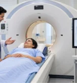
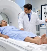
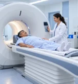
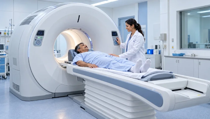
You lie on the scanning table while the scanner obtains the PET and CT images. It is important to remain still during image acquisition.
5. Review and reporting
The images are assessed by an appropriately trained specialist, and the report is sent to the referring doctor for interpretation in the context of your clinical history.
The injection may cause brief discomfort at the injection site. The imaging itself is generally painless, although remaining still on the table may cause some discomfort.
Is the Radioactive Tracer in PET-CT Safe?
PET-CT uses a controlled amount of radioactive tracer for diagnostic imaging. The tracer’s radioactivity decreases over time, and the substance is eliminated from the body through natural processes, including urine.
However, PET-CT involves exposure to ionizing radiation from the radiotracer, and the CT component also uses X-rays. The examination should therefore be performed when there is an appropriate medical reason.
Tell your doctor and imaging team if you are pregnant, may be pregnant or are breastfeeding. They can explain the relevant precautions and whether the planned examination is appropriate.
Follow any after-scan instructions provided by the imaging team. These may vary depending on the tracer and examination.
What Do PET-CT Scan Results Mean?
PET-CT images show patterns of tracer uptake alongside anatomical information from CT. These findings can help doctors assess a suspected condition, but the results must be interpreted carefully.
Increased tracer uptake does not automatically confirm cancer. Infection, inflammation and other processes may also cause increased uptake. Conversely, some tumours may not show the expected pattern of activity on a particular PET examination.
The radiology or nuclear medicine report is considered alongside symptoms, previous imaging, laboratory results, biopsy findings and other relevant clinical information. Additional tests may sometimes be needed before a diagnosis or treatment decision can be made.
Your treating doctor can explain what the findings mean for your individual situation.
Frequently Asked Questions (FAQs)
1. Does being advised a PET-CT scan mean I have cancer?
No. A PET-CT scan can be recommended for several reasons, including evaluating a known condition or investigating a specific clinical question. The recommendation alone does not establish a cancer diagnosis.
2. Is a PET-CT scan painful?
The imaging itself is generally painless. You may feel brief discomfort when the tracer is injected, and you will need to remain still while the images are obtained.
3. Can infection cause increased uptake on a PET-CT scan?
Yes. Infection and inflammation can cause increased FDG uptake. The findings must be interpreted with the CT images, medical history and other investigations.
4. Can I eat before a PET-CT scan?
For many FDG PET-CT examinations, fasting is required for several hours. Instructions vary, so follow the exact preparation guidance from your imaging centre. Patients with diabetes should request individual instructions.
5. What is the difference between FDG, PSMA and DOTA PET imaging?
They use different tracers for different biological targets and clinical purposes. FDG is widely used for many cancers, PSMA is used in appropriate prostate cancer assessments, and DOTA-based tracers are used for selected neuroendocrine tumours. The correct choice depends on the medical indication.
6. How long does a PET-CT scan take?
The total appointment includes preparation, tracer injection, a resting period and image acquisition. The time varies according to the tracer and protocol, so confirm the expected duration with the imaging centre.
7. Where can I enquire about a PET-CT scan in Surat?
You can contact Super Scan to enquire about PET-CT services at its Khatodara branch, including the examination required, tracer availability, appointment arrangements and preparation instructions.
Also read,
- Diagnostic Imaging Services in Surat
- Colour Doppler
- Contrast Procedures
- 3D and 4D Sonography
- PET Scan
- Gamma Scan