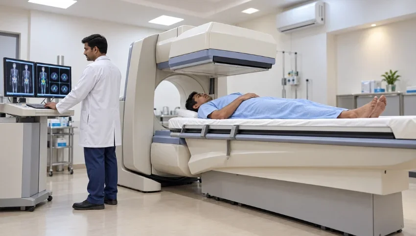
Understand how Gamma Scan and gamma camera imaging work, when nuclear medicine scans may be recommended, how the procedure is performed, and what patients should know before the examination.
Advanced Nuclear Imaging to Understand How the Body Functions.
What Is a Gamma Scan?
A Gamma Scan is a type of nuclear medicine imaging examination that uses a small amount of radioactive material, called a radiotracer or radiopharmaceutical, to provide information about how specific organs, tissues or body systems are functioning.
Unlike conventional imaging methods that primarily show anatomy, nuclear medicine can provide functional or physiological information. After the radiotracer reaches the area being examined, a specialized gamma camera detects the radiation emitted by the tracer and uses this information to create images. The gamma camera itself does not emit radiation.
Depending on the clinical question, different nuclear medicine examinations may be performed using a gamma camera. These can include selected bone, thyroid, kidney, hepatobiliary and cardiac studies, among others.
How Does a Gamma Camera Scan Work?
A Gamma Scan generally involves administering a carefully selected radiotracer into the body. Depending on the examination, the tracer may be injected, swallowed or administered in another appropriate way.
The radiotracer travels to the area being studied and accumulates according to the biological process or tissue being evaluated. The gamma camera then detects the energy emitted by the radiotracer and a computer processes the information to create images.
The timing of imaging can vary considerably depending on the type of nuclear medicine examination. Some scans begin shortly after administration of the tracer, while others require a waiting period before images are obtained.
What Can a Gamma Scan Be Used For?
The specific purpose of a Gamma Scan depends on the type of nuclear medicine study prescribed by the doctor.
Gamma camera imaging may be used to evaluate different organs and physiological processes, including:
- Bone activity and selected bone conditions
- Thyroid function and uptake
- Kidney function and drainage
- Liver, gallbladder and bile-duct function
- Heart blood-flow and selected cardiac conditions
- Certain areas of inflammation or abnormal biological activity
- Selected conditions where functional information is important
Nuclear medicine examinations can provide information that may not be available from anatomical imaging alone.
The exact scan should always be selected according to the patient’s symptoms, medical history and the clinical question that needs to be answered.
Gamma Scan for Bone Evaluation
A nuclear medicine bone scan is one example of imaging performed using a gamma camera.
It can help evaluate changes in bone activity and may be considered in selected situations involving unexplained bone pain, suspected bone abnormalities, certain infections, injuries or cancer-related assessment.
A bone scan does not simply show the shape of a bone. It provides information about areas of increased or altered bone activity.
The interpretation of these findings must be considered together with the patient’s symptoms, medical history and other imaging or laboratory findings.
Gamma Scan for Thyroid Evaluation
Certain thyroid nuclear medicine examinations use a radiotracer and gamma camera to evaluate thyroid activity and tracer uptake.
The examination may help doctors assess selected thyroid conditions and understand how different areas of the thyroid are functioning.
The preparation and type of tracer depend on the specific thyroid examination being performed. Your doctor or nuclear medicine team will provide the appropriate instructions before the scan.
Gamma Scan for Kidney Function
Some nuclear medicine kidney examinations, also known as renal scintigraphy, can provide information about kidney function, blood flow and urinary drainage.
This can be different from a conventional anatomical examination because nuclear medicine can provide functional information about how the kidneys are working.
The exact protocol depends on the clinical indication and the information required by the referring doctor.
Gamma Scan for Liver and Gallbladder Evaluation
Certain hepatobiliary nuclear medicine examinations can evaluate the liver, gallbladder and bile ducts.
A radiotracer travels through the relevant system, and the gamma camera records its movement and distribution over time. This can provide functional information that may complement ultrasound, CT or MRI findings.
Gamma Scan for Heart Evaluation
Nuclear medicine can also be used for selected cardiac examinations.
Cardiac nuclear medicine imaging may help evaluate blood flow to the heart muscle and certain heart conditions. In some studies, images are obtained during stress and at rest so that doctors can compare blood flow under different conditions.
The appropriate cardiac nuclear medicine examination depends on the patient’s symptoms, medical history and the clinical question.
Is Gamma Scan the Same as PET-CT?
No. Gamma camera imaging and PET-CT are both forms of nuclear medicine, but they use different imaging technologies and radiotracers.
A conventional gamma camera detects gamma radiation from certain radiotracers and can produce planar or SPECT images. SPECT uses rotating gamma-camera detectors to create more detailed three-dimensional images.
PET-CT uses PET technology together with CT and is designed to provide metabolic information alongside anatomical information.
Therefore, a Gamma Scan, SPECT, SPECT/CT and PET-CT are not interchangeable examinations. The appropriate study depends on the disease being investigated and the information required.
What Is SPECT?
SPECT stands for Single Photon Emission Computed Tomography.
In SPECT imaging, gamma-camera detectors rotate around the patient’s body to acquire multiple images. A computer then reconstructs the information into detailed three-dimensional images.
SPECT can provide additional information compared with conventional planar nuclear medicine imaging and may also be combined with CT as SPECT/CT.
Is a Gamma Scan Safe?
Nuclear medicine examinations use a small amount of radioactive material selected for the particular examination.
As with any medical procedure involving radiation, the examination should be performed when it is clinically appropriate. The amount and type of radiotracer depend on the specific nuclear medicine study.
The radioactive material naturally loses radioactivity over time, and depending on the tracer, some of it may leave the body through urine or stool. Patients receive specific instructions when additional precautions or preparation are required.
Patients should tell their doctor and nuclear medicine team if they are pregnant or breastfeeding before the examination. They should also provide information about medications, allergies, recent illnesses and relevant medical conditions.
Does a Gamma Camera Emit Radiation?
No.
The gamma camera detects radiation emitted by the radiotracer that has been administered to the patient. The camera itself does not produce the radiation used to create the nuclear medicine images.
This is an important distinction between the radioactive tracer used during the examination and the imaging equipment that detects the emitted energy.
What Happens During a Gamma Scan?
The exact procedure varies according to the type of scan, but the process may generally involve:
- Medical assessment: The nuclear medicine team reviews the examination and relevant patient information.
- Radiotracer administration: The prescribed radiotracer may be injected, swallowed or administered according to the examination protocol.
- Waiting period: Some examinations require time for the tracer to reach and concentrate in the area being studied.
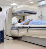
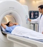
- Image acquisition: The patient lies on an examination table while the gamma camera records images.
- Additional views: Depending on the examination, different positions or additional images may be required.
- Image interpretation: A trained specialist reviews the images and prepares a report for the referring doctor.
Patients generally need to remain still during image acquisition to help obtain clear images.
How Should You Prepare for a Gamma Scan?
Preparation depends on the type of nuclear medicine examination.
Your doctor or imaging team may provide instructions regarding food, drinks, medicines or other preparation. Patients should not assume that every Gamma Scan requires the same preparation.
Before the examination, inform the medical team about:
- Pregnancy or possible pregnancy
- Breastfeeding
- Current medications
- Allergies
- Recent illnesses
- Relevant medical conditions
- Previous examinations or treatments when requested
Jewellery and certain accessories may need to be removed because they can interfere with imaging.
How Long Does a Gamma Scan Take?
There is no single duration for every Gamma Scan.
The time depends on the specific nuclear medicine examination, the radiotracer used and how long the tracer needs to reach the area being evaluated.
Some examinations can involve imaging shortly after tracer administration, while others require one or more waiting periods and multiple imaging sessions.
Your nuclear medicine team can explain the expected timing before the examination.
What Is the Difference Between Gamma Scan and CT or MRI?
Gamma Scan, CT and MRI provide different types of information.
Gamma Scan: Primarily provides functional information related to the distribution and activity of a radiotracer.
CT: Uses X-rays to provide detailed anatomical cross-sectional images.
MRI: Uses a strong magnetic field and radiofrequency signals to provide detailed images, particularly of many soft tissues, without ionizing radiation.
This means that a Gamma Scan is not necessarily an alternative to CT or MRI. In some clinical situations, different imaging examinations may complement one another.
When Might a Doctor Recommend Nuclear Medicine Imaging?
A doctor may recommend a nuclear medicine examination when information about organ function, blood flow, metabolism or another physiological process is important to the diagnosis.
The decision depends on:
- Symptoms
- Medical history
- Physical examination
- Suspected condition
- Previous imaging results
- Laboratory findings
- The specific clinical question
Patients should avoid choosing a nuclear medicine scan solely because it appears more advanced than another imaging test.
The right examination is the one that provides the information needed for the patient’s particular medical situation.
Why Choosing the Right Nuclear Medicine Examination Matters
“Gamma Scan” is a broad term from a patient’s perspective, but nuclear medicine includes several different examinations with different purposes.
A thyroid scan is not the same as a bone scan. A renal scan is not the same as a cardiac nuclear medicine study. SPECT may provide three-dimensional functional imaging, while SPECT/CT combines functional information with anatomical CT imaging.
Therefore, the important question is not simply:
“Do I need a Gamma Scan?”
It is:
“Which nuclear medicine examination is appropriate for my medical condition?”
Your doctor and nuclear medicine specialist can determine the appropriate test based on your individual clinical situation.
Frequently Asked Questions
1. What is a Gamma Scan?
A Gamma Scan is a nuclear medicine imaging examination in which a gamma camera detects radiation emitted by a radiotracer to create images and provide functional information about selected organs or tissues.
2. Is Gamma Scan the same as a Gamma Camera Scan?
The terms may be used interchangeably by patients, but technically the gamma camera is the imaging equipment used to detect radiation from the radiotracer and produce the images.
3. Does a Gamma Scan use radiation?
Yes. Nuclear medicine imaging involves a radiotracer that emits radiation, which is detected by the gamma camera. The amount and type of radiotracer depend on the specific examination.
4. Does the Gamma Camera itself emit radiation?
No. The gamma camera detects radiation emitted from the radiotracer administered to the patient.
5. Is Gamma Scan painful?
The imaging portion is generally painless. If an injection is required, the patient may experience the brief discomfort associated with an injection.
6. Can Gamma Scan detect cancer?
Some nuclear medicine examinations can help evaluate certain cancers or cancer-related conditions, but the appropriate examination depends on the type of cancer and the clinical question. Nuclear medicine is also used for many non-cancer conditions.
7. Can Gamma Scan be used for thyroid problems?
Yes. Certain thyroid nuclear medicine examinations use a radiotracer and gamma camera to evaluate thyroid activity and tracer uptake.
8. Is Gamma Scan the same as PET-CT?
No. Both are nuclear medicine techniques, but they use different technologies and radiotracers. PET-CT combines PET imaging with CT, while gamma-camera studies may use planar imaging or SPECT/SPECT-CT.
9. Can I eat before a Gamma Scan?
Preparation varies by examination. Follow the specific instructions provided by your doctor or nuclear medicine team rather than assuming that every Gamma Scan requires fasting.