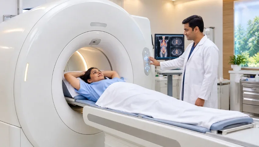
Understand the differences between MRI, CT, PET-CT and ultrasound and why the right imaging test depends on your symptoms, medical condition and clinical question.
The right scan is the one that answers the right medical question.
When a doctor recommends an imaging test, patients often wonder why one person is advised an ultrasound while another may need a CT scan, MRI or PET-CT. The answer is that MRI vs CT vs PET-CT vs ultrasound is not really a competition between four technologies. Each imaging method provides different information and is useful for different medical situations.
There is no single imaging scan that is universally best. The appropriate examination depends on your symptoms, medical history, physical examination, previous test results and the specific medical question your doctor needs answered.
Let us understand the differences in simple terms.
Ultrasound: A Common First-Line Imaging Test
Ultrasound, also called sonography, uses high-frequency sound waves to create images of structures inside the body. It does not use ionizing radiation and is widely used for evaluating many organs and conditions.
Ultrasound may be useful for examining:
- Liver and gallbladder
- Kidneys and urinary tract
- Uterus and ovaries
- Thyroid gland
- Breast and soft-tissue lumps
- Abdominal organs
- Pregnancy-related conditions
- Blood flow through arteries and veins with Doppler ultrasound
Because ultrasound is widely available and does not use ionizing radiation, it is often used as an initial examination when clinically appropriate.
Why Isn’t Ultrasound Used for Everything?
Ultrasound has important limitations. Sound waves do not travel effectively through air and may have difficulty passing through bone. Image quality can also vary depending on the body region being examined and individual patient factors.
For some conditions, an ultrasound may identify an abnormality but cannot fully characterize it. In such cases, the doctor may recommend CT or MRI for additional information.
This does not necessarily mean that the ultrasound was inadequate. The second examination may simply be answering a different medical question.
CT Scan: Fast and Detailed Imaging
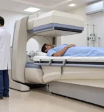
Computed tomography, commonly called a CT scan, uses X-rays and computer processing to create detailed cross-sectional images of the body.
One of the major advantages of CT is speed. A modern CT examination can evaluate many parts of the body rapidly, making it particularly valuable when urgent information is needed.
CT may be used for:
- Emergency and trauma assessment
- Chest and lung evaluation
- Abdominal conditions
- Kidney stones
- Bone injuries and abnormalities
- Internal bleeding assessment
- Certain vascular examinations
- Treatment planning and follow-up in selected conditions
CT can also be combined with contrast material when additional visualization of certain organs, tissues or blood vessels is required.
Does CT Scan Use Radiation?
Yes. CT uses X-rays, which are a form of ionizing radiation. The amount of radiation varies according to the examination and patient factors.
However, a medically necessary CT examination should not be avoided simply because it uses radiation. The FDA notes that when CT is appropriately used, its diagnostic benefits generally outweigh the small radiation-related risk, while unnecessary exposure should be minimized.
The important question is therefore not simply:
“Does CT use radiation?”
It is:
“Is CT the appropriate examination to answer the medical question?”
Your treating doctor and radiologist can consider the potential benefits, risks and available alternatives before selecting the examination.
MRI: Detailed Soft-Tissue Imaging Without Ionizing Radiation
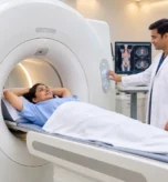
Magnetic resonance imaging, or MRI, uses a strong magnetic field and radiofrequency signals to produce detailed images of the body’s internal structures. MRI does not use ionizing radiation.
MRI can be particularly useful for evaluating soft tissues and may be recommended for areas such as:
- Brain and nervous system
- Spine
- Knees, shoulders and other joints
- Muscles, tendons and ligaments
- Liver and selected abdominal organs
- Pelvis
- Prostate
- Breast
For example, when a knee injury raises concern about a ligament or cartilage problem, MRI can provide detailed information about structures that may not be adequately evaluated with other imaging methods.
MRI can also provide detailed assessment of many neurological and spinal conditions.
Why Not Use MRI for Every Patient?
MRI is not automatically the best choice for every condition.
CT can be considerably faster and may be more appropriate in many emergency situations. CT can also be particularly useful for evaluating the lungs, trauma, kidney stones and certain bone abnormalities.
MRI examinations may take longer and require the patient to remain still. MRI safety screening is also important because the examination uses a powerful magnetic field.
Patients should inform the imaging team about relevant implanted devices, metallic objects, previous surgeries involving metal, or any other potential MRI safety concerns before the examination.
PET-CT: Combining Anatomy With Metabolic Information
PET-CT provides a different type of information from conventional CT or MRI.
A PET examination uses a radiotracer to provide information about biological or metabolic activity, while the CT component provides anatomical information. The two sets of information are combined to help doctors assess certain diseases.
PET-CT has important applications in cancer care, where it may be used in selected patients to help:
- Assess the extent of disease
- Stage certain cancers
- Evaluate treatment response
- Investigate suspected recurrence
- Identify metabolically active areas that may require further evaluation
Different PET tracers are designed for different clinical purposes.
For example, FDG PET-CT is commonly used in the evaluation of many cancers, while specialized tracers such as PSMA or DOTA-based tracers may be used for selected prostate cancers or neuroendocrine tumors, respectively, when clinically appropriate.
Therefore, even the question “Do I need a PET-CT?” may need to be followed by another question:
“Which PET-CT examination is appropriate for my condition?”
MRI, CT, PET-CT or Ultrasound: Which Is Used for Cancer?
Patients with cancer often ask which scan is best for cancer diagnosis or follow-up.
There is no single answer because cancer imaging depends on the type of cancer, its location, the clinical situation and the information doctors need.
Depending on the situation, doctors may use:
- Ultrasound
- Mammography
- CT
- MRI
- PET-CT
- A combination of imaging examinations
For example, breast cancer evaluation may involve mammography and ultrasound, with breast MRI used in selected situations.
Prostate cancer evaluation may involve prostate MRI and, when appropriate, PSMA PET-CT.
Other cancers may require CT, MRI, ultrasound or FDG PET-CT depending on the clinical question.
These examinations are not necessarily competing with one another. They can provide complementary information.
What Is the Role of Whole-Body MRI?
Whole-body MRI can examine multiple regions of the body without ionizing radiation and has important applications in selected clinical situations.
However, it should not be considered a universal replacement for every other imaging examination.
Different imaging technologies have different strengths and limitations. A whole-body MRI cannot guarantee detection of every disease or every early cancer.
The appropriate imaging strategy should therefore be based on the patient’s symptoms, age, risk factors, medical history and clinical indication rather than simply choosing the most advanced-sounding scan.
Why Might I Need a Second Scan After an Ultrasound or CT?
Patients sometimes become concerned when their doctor recommends another imaging examination after an initial scan.
For example:
“My ultrasound showed something. Why do I now need an MRI?”
This does not necessarily mean that the first examination failed.
An initial examination may identify an area that needs more detailed characterization. A different imaging technique may then provide information that the first examination was not designed to provide.
For instance, an ultrasound may identify a liver lesion, after which MRI may be recommended in an appropriate clinical setting to characterize the lesion more accurately.
The second examination may therefore be the next step in answering the clinical question.
Can You Choose Your Own Imaging Scan?
It is generally better not to select an imaging examination simply because one technology sounds more advanced or expensive.
The appropriate scan depends on factors such as:
- Symptoms
- Age
- Medical history
- Physical examination
- Previous imaging results
- Suspected condition
- Body region being evaluated
- The specific clinical question
Your treating doctor and radiologist can determine which imaging examination is most likely to provide useful diagnostic information.
Sometimes a simple ultrasound may be sufficient.
In another situation, MRI, CT or PET-CT may be more appropriate.
More advanced or more expensive does not automatically mean more appropriate.
A Simple Way to Understand the Four Imaging Tests
A simple comparison can help:
Ultrasound
Useful for many abdominal and pelvic organs, pregnancy-related imaging, lumps and swellings, and blood-flow assessment with Doppler. It does not use ionizing radiation.
CT Scan
Provides fast, detailed cross-sectional images and is particularly useful in many emergency situations, trauma assessments, chest and abdominal examinations, kidney stones and selected vascular studies.
MRI
Provides excellent soft-tissue detail without ionizing radiation and is widely used for the brain, spine, joints, muscles, ligaments and selected abdominal, pelvic, prostate and breast examinations.
PET-CT
Combines metabolic information from PET with anatomical information from CT and has important applications in selected cancer evaluations and other specialized clinical situations.
None of these technologies is universally superior. They are different diagnostic tools designed to answer different medical questions.
Choosing the Right Imaging Test in Surat
For patients in Surat and South Gujarat, access to different imaging technologies can be important because different clinical situations may require different examinations.
MRI, CT, PET-CT, ultrasound and other diagnostic imaging techniques each have their own role. The goal should not simply be to choose the most sophisticated scan. The goal is to select the examination that can provide the information needed for the patient’s particular medical problem.
For patients being evaluated for cancer or other complex conditions, imaging may sometimes involve more than one examination. The treating doctor and radiologist can review the clinical history and previous findings to determine what additional information is required.
Patients should therefore avoid self-selecting an imaging examination based only on online information or the perceived sophistication of a technology.
Frequently Asked Questions
Is MRI better than CT?
Not necessarily. MRI and CT are designed for different clinical purposes. MRI provides excellent soft-tissue detail without ionizing radiation, while CT is fast and highly useful for many emergency, chest, abdominal, trauma and bone-related evaluations.
Is ultrasound safer than CT?
Ultrasound does not use ionizing radiation, whereas CT uses X-rays. However, the appropriate examination depends on the clinical question. A medically necessary CT may provide information that ultrasound cannot.
Does MRI use radiation?
MRI does not use ionizing radiation. It uses a strong magnetic field and radiofrequency signals to generate images.
Why is PET-CT used in cancer?
PET-CT can provide information about both anatomy and biological activity. In selected cancers, this may help doctors assess disease extent, treatment response or suspected recurrence. The appropriate tracer depends on the disease and clinical indication.
Can one scan detect every disease?
No. Every imaging technique has strengths and limitations. Different examinations may be used separately or together depending on the clinical question.
Can I request an MRI instead of CT?
You can discuss your preferences and concerns with your doctor, but the final imaging choice should be based on the medical question, patient factors and the information required for diagnosis.
Why do I sometimes need more than one imaging test?
Different imaging techniques provide different types of information. A second examination may be recommended to further characterize a finding or answer a question that the first scan could not fully address.