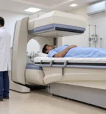
Ultrasound Scan in Surat: Complete USG Imaging Guide
An Ultrasound Scan is a non-invasive diagnostic imaging examination that uses high-frequency sound waves to create real-time images of structures inside the body. Unlike X-ray and CT imaging, ultrasound does not use ionizing radiation. It can be used to evaluate many organs, soft tissues, blood vessels and, when medically indicated, pregnancy-related structures.
For patients searching for an Ultrasound Scan in Surat, understanding what USG is, why doctors recommend it, how different examinations work, how to prepare and what to expect during the appointment can make the diagnostic process easier and less stressful.
What Is an Ultrasound Scan?
Ultrasound, commonly called USG or sonography, uses a handheld device called a transducer. The transducer sends high-frequency sound waves into the body and receives the returning echoes. A computer processes these echoes to create images that can be viewed on a monitor in real time.
Depending on the examination, ultrasound can provide information about the size, shape, structure and movement of organs and tissues. It can also be used to assess blood flow through Doppler techniques.
How Does Ultrasound Imaging Work?
During a typical examination, a sonographer or radiologist applies a water-based gel to the skin over the area being examined. The transducer is then gently moved across the skin.
The gel helps create proper contact between the transducer and the skin, allowing sound waves to travel efficiently into the body. Returning echoes are converted into images that appear on the screen in real time.
Most routine examinations are well tolerated, although pressure from the transducer may cause temporary discomfort if the area being examined is already tender.
What Can an Ultrasound Scan Detect?
USG imaging can be used for many diagnostic purposes. Depending on the clinical requirement, it can help doctors evaluate:
- Liver and gallbladder
- Kidneys and urinary bladder
- Pancreas and spleen
- Thyroid and other superficial structures
- Uterus and ovaries
- Pregnancy and fetal development
- Breast abnormalities
- Muscles, tendons and selected soft tissues
- Blood vessels and blood flow
- Certain abdominal and pelvic conditions
Ultrasound is particularly useful for evaluating soft tissues and fluid-filled structures and can also provide real-time imaging during certain procedures.
Common Types of Ultrasound Scans
The exact examination depends on the patient’s symptoms and the doctor’s recommendation.
Abdominal Ultrasound
An abdominal ultrasound may be recommended to evaluate organs such as the liver, gallbladder, kidneys, pancreas, spleen and abdominal blood vessels. It may be requested for symptoms such as abdominal pain, swelling or other suspected abnormalities.
Pelvic Ultrasound
Pelvic imaging can evaluate structures such as the uterus, ovaries and urinary bladder. It may be recommended for pelvic pain, abnormal bleeding, menstrual concerns or other gynecological conditions.
Pregnancy Ultrasound
Ultrasound is commonly used during pregnancy to monitor the developing fetus and evaluate pregnancy-related structures. It does not use ionizing radiation and is widely used when medically indicated during pregnancy.
However, pregnancy ultrasound should be performed for appropriate medical reasons and according to the advice of the treating healthcare professional.
Breast Ultrasound
Breast ultrasound uses sound waves to create images of breast tissue. It can help evaluate a breast lump or an abnormality detected during a physical examination or another imaging examination.
Thyroid Ultrasound
A thyroid ultrasound can provide detailed images of the thyroid gland and help evaluate nodules, enlargement and other structural abnormalities.
Kidney and Urinary Ultrasound
Renal or urinary ultrasound can be used to evaluate the kidneys and urinary bladder. Depending on the clinical indication, it may help assess structural abnormalities, swelling, stones or other concerns.
Musculoskeletal Ultrasound
Musculoskeletal ultrasound can evaluate selected muscles, tendons, ligaments and other superficial soft tissues. It can also provide real-time information about movement in certain situations.
Vascular Ultrasound
Vascular ultrasound uses sound waves to evaluate blood vessels. Doppler techniques can help assess blood flow and identify problems such as narrowing or blood clots in selected vessels.
What Is Doppler Ultrasound?
Doppler Ultrasound is a specialized ultrasound technique used to evaluate the direction and speed of blood flow through blood vessels.
The movement of blood cells changes the frequency of the reflected sound waves. The ultrasound system processes these changes to provide information about blood flow, which can be displayed as waveforms, measurements or colour images depending on the technique.
Doppler studies may be recommended for evaluating arteries, veins and blood flow to selected organs.
What Are the Benefits of Ultrasound?
Ultrasound has several characteristics that make it useful for diagnostic imaging:
No Ionizing Radiation
Ultrasound uses sound waves rather than X-rays, so it does not expose the patient to ionizing radiation.
Real-Time Imaging
The images are generated in real time, allowing movement and certain physiological processes to be observed during the examination.
Non-Invasive Examination
Most external ultrasound examinations do not require needles, injections or surgical procedures.
Useful for Soft Tissues
Ultrasound can provide useful images of soft tissues that may not be clearly demonstrated on conventional X-rays.
Widely Used During Pregnancy
When medically indicated, ultrasound is commonly used to monitor pregnancy and fetal development.
Is an Ultrasound Scan Safe?
Diagnostic ultrasound is generally considered safe when performed appropriately by trained healthcare professionals. It uses non-ionizing sound waves rather than X-rays.
However, ultrasound energy can produce biological effects under certain conditions, so medical organizations recommend prudent use, particularly during pregnancy. Ultrasound should therefore be performed for appropriate clinical reasons rather than solely for non-medical purposes.
How Should You Prepare for an Ultrasound Scan?
Preparation depends on the body part being examined.
For some examinations, you may be asked to avoid food or drinks for a specific period. For others, such as certain pelvic or urinary examinations, you may be asked to drink water and maintain a full bladder before the scan.
General preparation may include:
- Wear comfortable, loose-fitting clothing.
- Follow the specific instructions given by the imaging centre.
- Avoid jewellery around the examination area when instructed.
- Bring your doctor’s prescription or referral.
- Carry previous relevant imaging reports when requested.
- Follow fasting instructions if your examination requires them.
- Follow bladder-filling instructions if applicable.
Do not follow a generic fasting or water-drinking rule for every USG examination. Preparation varies according to the specific scan.
What Happens During an Ultrasound Examination?
You will generally lie on an examination table. The sonographer or radiologist may ask you to change position depending on the area being examined.
A small amount of gel is applied to the skin. The transducer is moved over the examination area to obtain the required images.
Most external ultrasound examinations are painless. Some pressure may be felt when the transducer is moved over a sensitive area.
After the examination, the gel is wiped away and most patients can return to their normal activities immediately.
How Long Does an Ultrasound Scan Take?
Many routine ultrasound examinations take around 30 minutes, although the duration can vary depending on the body part, examination type and complexity. Some more extensive examinations may take longer.
Your imaging centre can provide more specific timing when your appointment is scheduled.
Ultrasound Scan for Pregnancy
Ultrasound plays an important role in pregnancy care because it can provide real-time images of the developing fetus without using ionizing radiation.
The timing and type of pregnancy ultrasound depend on the pregnancy stage and the treating doctor’s recommendation.
Patients should remember that an ultrasound report is interpreted in the context of the pregnancy history, symptoms and other clinical information. Your obstetrician or healthcare provider should explain what the findings mean for your individual situation.
Choosing an Ultrasound Centre in Surat
When selecting a diagnostic imaging centre, patients should consider:
- Appropriate ultrasound technology
- Experienced radiology professionals
- Qualified sonography staff
- Patient safety and privacy
- Appropriate examination protocols
- Clear preparation instructions
- Reliable reporting
- Convenient appointment scheduling
- Availability of previous reports for comparison when clinically useful
The quality of a diagnostic examination depends not only on the equipment but also on appropriate protocols, patient cooperation and professional interpretation.
Frequently Asked Questions About Ultrasound
Is an ultrasound scan painful?
Most external ultrasound examinations are painless. Some patients may feel temporary pressure, especially when the examination area is already tender.
Does ultrasound use radiation?
No. Ultrasound uses high-frequency sound waves and does not use ionizing radiation.
Can ultrasound be performed during pregnancy?
Ultrasound is commonly used during pregnancy when medically indicated and does not use ionizing radiation.
Do I need to fast before an ultrasound?
Not necessarily. Preparation depends on the type of examination. Some scans may require fasting, while others may require drinking water and having a full bladder.
How long does an ultrasound take?
Many routine examinations take around 30 minutes, but the actual duration depends on the type and complexity of the examination.
What is the difference between USG and ultrasound?
USG, ultrasound and sonography are commonly used terms for ultrasound imaging. The exact examination performed depends on the body area and clinical requirement.
Is Doppler the same as a regular ultrasound?
Doppler is a specialized ultrasound technique that evaluates blood flow through vessels. It may be performed as part of a broader ultrasound examination or as a dedicated vascular study.
Can ultrasound detect every disease?
No. Ultrasound has important capabilities but also limitations. Some body structures are difficult to evaluate with ultrasound, and other imaging methods such as CT or MRI may be recommended depending on the clinical question.