160 Slice CT Scan in Surat: Advanced CT Imaging
160 Slice CT Scan is an advanced multislice computed tomography technique used to create detailed cross-sectional images of the body. CT combines X-ray technology with computer processing to produce images of organs, bones, soft tissues and blood vessels. These images can also be reconstructed in different planes and, when appropriate, into three-dimensional views, helping doctors and radiologists evaluate a wide range of medical conditions.
For patients looking for advanced CT scan services in Surat, understanding how multislice CT works, when it is recommended, how to prepare and what safety precautions are involved can make the diagnostic process easier to understand.
What Is a 160 Slice CT Scan?
A 160 Slice CT Scan is a type of multislice CT technology designed to acquire multiple image slices during each rotation of the scanner. The resulting data can be processed to create detailed images of different anatomical regions.
CT imaging can be used to examine the head, chest, abdomen, pelvis, bones, blood vessels and other structures depending on the clinical requirement. The exact CT protocol is selected according to the body part being evaluated and the diagnostic question that needs to be answered.
How Does a 160 Slice CT Scan Work?
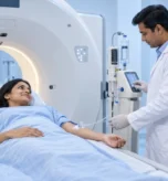
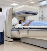
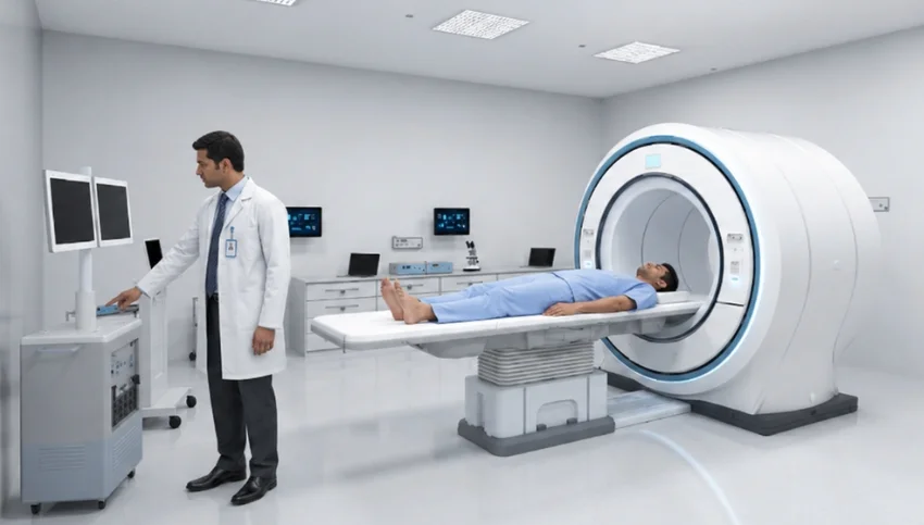
During the examination, the patient lies on a motorized table that moves through the CT scanner’s circular opening. X-ray components rotate around the body while detectors collect imaging information.
A computer then processes the information to create cross-sectional images. These images can be viewed in multiple planes and may also be used to generate 3D reconstructions when clinically appropriate.
The examination is generally quick, and patients are required to remain as still as possible during image acquisition to help maintain image quality.
Why Is Multislice CT Used?
Multislice CT can provide detailed images quickly, making CT useful for a wide range of diagnostic situations. It can help evaluate bones, internal organs, soft tissues and blood vessels within the same examination.
CT is also commonly used in emergency situations because it can rapidly demonstrate internal injuries, bleeding and other abnormalities that may require urgent medical attention.
What Conditions Can a CT Scan Help Evaluate?
The examination recommended by your doctor will depend on your symptoms and clinical history.
Head and Brain CT
A head CT can help evaluate certain injuries, bleeding, structural abnormalities and other neurological concerns. The appropriate CT protocol depends on the patient’s clinical situation.
Chest CT
Chest CT can provide detailed images of the lungs, airways, blood vessels and other structures within the chest. It may be recommended for unexplained respiratory symptoms or to further evaluate abnormalities identified through other investigations.
Abdominal and Pelvic CT
Abdominal and pelvic CT may be recommended for conditions such as abdominal pain, suspected kidney stones, appendicitis, diverticulitis and evaluation of various abdominal or pelvic abnormalities.
Bone and Musculoskeletal Imaging
CT provides detailed information about bones and can be useful for evaluating fractures, complex skeletal structures and selected orthopedic conditions.
Blood Vessel Imaging
Specialized CT techniques can be used to examine blood vessels and provide detailed information about vascular structures when clinically indicated.
What Are the Benefits of Advanced CT Imaging?
Depending on the examination, CT can offer several important diagnostic advantages:
- Detailed cross-sectional images
- Rapid image acquisition
- Evaluation of bones and soft tissues
- Assessment of blood vessels with specialized protocols
- Multiplanar image reconstruction
- Possible 3D image reconstruction
- Useful imaging in emergency situations
CT is considered a fast, non-invasive and accurate diagnostic imaging method for many clinical applications.
However, the benefit of any CT examination depends on selecting the appropriate protocol for the patient’s clinical requirement.
160 Slice CT Scan vs Conventional CT
The term “160 slice” describes the detector configuration and image acquisition capabilities of a multislice CT system. Modern multislice CT technology can acquire multiple sections during a rotation and process the resulting information into detailed images.
However, the number of slices alone should not be treated as the only measure of diagnostic quality. Image quality can also depend on the scanner, acquisition protocol, patient movement, reconstruction techniques, radiation-dose management and the expertise involved in interpreting the examination.
For this reason, patients should focus on whether the imaging centre can provide the specific CT examination recommended by their doctor rather than choosing a centre solely on the basis of slice count.
Is a 160 Slice CT Scan Safe?
CT uses X-rays, so it involves exposure to ionizing radiation. The amount of radiation varies according to the examination and protocol.
Radiology professionals aim to use the lowest radiation exposure reasonably necessary to obtain diagnostically useful images. When a CT examination is medically justified, its diagnostic benefits generally outweigh the small potential radiation risk.
Patients should tell their doctor or CT technologist if there is a possibility of pregnancy. CT is generally avoided during pregnancy unless the examination is medically necessary.
Is Contrast Required for a 160 Slice CT Scan?
Contrast is not required for every CT scan.
Depending on the body part and the clinical question, a CT examination may be performed without contrast or with an appropriate contrast agent.
Iodine-based contrast material is commonly used for certain CT examinations because it can improve the visibility of selected organs, blood vessels and tissues.
If contrast is planned, patients should inform the medical team about:
- Previous reactions to contrast material
- Kidney disease
- Diabetes
- Thyroid problems
- Heart disease
- Known allergies
- Pregnancy or possible pregnancy
Serious reactions to iodinated contrast are uncommon, but appropriate patient screening is important.
How to Prepare for a 160 Slice CT Scan?
Preparation depends on the type of examination.
Before your appointment, you may be advised to:
- Follow the fasting instructions provided by the imaging centre.
- Bring your doctor’s prescription or referral.
- Carry previous CT, MRI, X-ray or ultrasound reports when relevant.
- Inform the team about medications and medical conditions.
- Mention any previous reaction to contrast material.
- Inform the team if you have kidney disease.
- Tell the staff if you are pregnant or may be pregnant.
- Remove jewellery or metallic objects when instructed.
For contrast-enhanced examinations, specific preparation instructions may be provided before the scan.
What Happens During the CT Examination?
You will usually lie on a narrow table that moves through the CT scanner. The technologist operates the scanner from a nearby control room.
You may be asked to hold your breath briefly during some examinations, particularly certain chest or abdominal scans. Remaining still is important because movement can reduce image quality.
If intravenous contrast is used, you may briefly experience a warm or flushed sensation or a metallic taste. These sensations generally pass quickly.
How Long Does a 160 Slice CT Scan Take?
The actual image acquisition for many CT examinations is relatively quick, although the complete appointment may take longer because of registration, patient preparation, positioning and, when required, contrast administration.
The duration varies according to the body region, examination protocol and whether contrast or additional imaging phases are required.
160 Slice CT Scan for Emergency Imaging
One of the important advantages of CT is its ability to provide diagnostic information quickly.
In emergency situations, CT may help doctors identify internal bleeding, traumatic injuries and other urgent abnormalities.
The exact imaging examination is determined by the emergency physician or referring doctor according to the patient’s condition.
Choosing an Advanced CT Scan Centre in Surat
When selecting a CT Scan Centre in Surat, consider more than the scanner’s slice count.
Important factors include:
- Appropriate CT technology
- Qualified radiology professionals
- Patient safety protocols
- Appropriate imaging protocols
- Contrast screening procedures
- Image quality and reporting
- Convenient appointment support
- Availability of previous imaging comparison when clinically useful
The right diagnostic centre should provide the examination recommended by your treating doctor while maintaining appropriate safety and reporting standards.
160 Slice CT Scan: Frequently Asked Questions
Is a 160 Slice CT Scan better than a regular CT scan?
A multislice system can acquire multiple image sections during a rotation and support detailed imaging. However, diagnostic quality depends on the complete imaging system, protocol, patient factors and interpretation—not simply the number of slices.
Does a 160 Slice CT Scan use radiation?
Yes. CT uses X-rays and therefore involves ionizing radiation. The radiation dose varies by examination, and imaging professionals aim to use an appropriate dose for the required diagnostic information.
Is contrast necessary for every CT scan?
No. Some CT examinations are performed without contrast, while others may require contrast to improve visualization of particular structures.
Can I have a CT scan if I have kidney disease?
Patients with kidney problems should inform the doctor and imaging team, particularly if contrast-enhanced CT is planned. The medical team can assess the potential benefits and risks before deciding whether contrast is appropriate.
Can pregnant women undergo CT?
Pregnancy or possible pregnancy should always be disclosed before a CT examination. Doctors generally avoid CT during pregnancy unless medically necessary because of potential radiation exposure to the developing baby.
How long does a CT scan take?
The image acquisition itself is often quick, but total appointment time varies according to the examination, preparation and whether contrast is required.
Can CT create 3D images?
Yes. CT data can be reformatted in different planes and can also be processed to create three-dimensional images when appropriate.