Understand how contrast-enhanced imaging improves the visibility of organs, tissues and body structures for more detailed diagnostic evaluation.
Contrast Procedures are specialized imaging examinations that use a contrast material to make selected organs, tissues, blood vessels or internal structures more clearly visible on medical images. Depending on the examination, contrast may be swallowed, administered through a vein, introduced into a specific body space or used during an image-guided procedure. At Super Scan, Contrast Procedures in Surat are performed under radiological guidance with appropriate patient-safety protocols to support accurate diagnostic evaluation.
Modern medical imaging can provide highly detailed pictures of the human body, but some organs and internal structures may be easier to evaluate when a suitable contrast material is used. Contrast Procedures are imaging examinations designed to improve the visibility of specific areas and help radiologists assess structures more clearly.
Contrast materials can be used with different imaging techniques, including X-ray, fluoroscopy, CT and MRI. Depending on the examination, they may be administered orally, intravenously, rectally or introduced into a specific body space. The appropriate method depends on the body part being examined and the clinical question being investigated.
For patients searching for Contrast Procedures in Surat, understanding the different examinations, preparation requirements and safety considerations can make the diagnostic process easier to understand.
What Are Contrast Procedures?
Contrast Procedures are diagnostic imaging examinations in which a contrast material is used to improve the visibility of particular organs, tissues, body cavities or other structures.
Contrast materials temporarily change how certain tissues or structures appear on medical images. They do not permanently stain organs and, depending on the type used, are generally eliminated from the body through normal biological processes.
The type of contrast material and method of administration depends on the imaging examination.
Common approaches include:
- Oral contrast
- Intravenous contrast
- Rectal contrast
- Contrast introduced into specific body spaces
- Contrast used during fluoroscopic examinations
The radiologist determines the appropriate examination and contrast technique according to the patient’s clinical requirements.
Why Are Contrast Procedures Performed?
The main purpose of contrast-enhanced imaging is to improve the visibility or differentiation of selected anatomical structures.
Contrast may help radiologists evaluate:
- The gastrointestinal tract
- Urinary structures
- Reproductive organs
- Blood vessels
- Certain organs and soft tissues
- Fistulas and abnormal passages
- Specific areas requiring image-guided evaluation
The exact purpose depends on the examination requested by the treating doctor.
Contrast is therefore not required for every imaging examination. A radiologist or referring doctor determines whether contrast is appropriate based on the patient’s symptoms, medical history and diagnostic question.
Types of Contrast Materials
Different examinations require different types of contrast materials.
Iodine-Based Contrast
Iodine-based contrast materials are commonly used with X-ray and CT examinations. They can also be administered into blood vessels during certain angiographic procedures.
When present in the area being examined, iodine-based contrast changes the appearance of tissues and blood vessels on X-ray-based images.
Barium-Based Contrast
Barium sulfate is commonly used for examinations of the gastrointestinal tract. It may be swallowed or administered rectally depending on the examination.
Barium-based contrast can help outline structures such as the esophagus, stomach, small intestine and colon on X-ray or fluoroscopic images.
Gadolinium-Based Contrast
Gadolinium-based contrast agents are commonly used for selected MRI examinations. They alter the magnetic properties of nearby water molecules, helping certain tissues and blood vessels appear differently on MRI images.
The decision to use gadolinium depends on the type of MRI examination and the information required.
Common Contrast Procedures at Super Scan
Super Scan’s dedicated Contrast Procedures service in Surat includes several specialized fluoroscopic contrast studies. These examinations are used to evaluate different parts of the digestive, urinary and reproductive systems.
Barium Swallow
A Barium Swallow is an imaging examination in which a patient drinks a barium-containing contrast material while X-ray or fluoroscopic images are obtained.
It can help evaluate the swallowing passage and upper gastrointestinal structures.
Barium Meal
A Barium Meal uses orally administered barium to outline parts of the upper gastrointestinal tract during imaging.
The examination may help the radiologist assess the appearance and movement of structures within the upper digestive system.
Follow Through
A Follow Through examination involves drinking contrast material followed by a series of abdominal images taken at intervals.
It is used to evaluate the passage of contrast through portions of the small intestine.
Barium Enema
A Barium Enema introduces contrast material through the rectum to outline the large intestine during imaging.
The examination allows the colon and rectum to be assessed under fluoroscopic or X-ray guidance.
HSG – Hysterosalpingography
Hysterosalpingography, commonly called HSG, is a specialized contrast examination of the uterus and fallopian tubes.
During the examination, contrast is introduced through the cervix while imaging is performed to evaluate the uterine cavity and fallopian tube pathways.
MCU – Micturating Cystourethrogram
MCU is a contrast-based fluoroscopic examination used to evaluate the urinary bladder and urethra during urination.
It may be recommended in selected clinical situations where the urinary tract needs dynamic imaging.
RGU – Retrograde Urethrogram
A Retrograde Urethrogram uses contrast introduced into the urethra while X-ray images are obtained.
It allows the urethra to be evaluated for abnormalities that may require further clinical assessment.
Sinogram
A Sinogram is an image-guided contrast examination of an abnormal tract or sinus.
Contrast is introduced into the tract, allowing its pathway and connection with surrounding structures to be visualized.
Fistulogram
A Fistulogram uses contrast imaging to evaluate a fistula, which is an abnormal connection or passage between body structures.
The contrast helps outline the pathway so that the radiologist can assess its extent and anatomical relationship.
How Is Contrast Administered?
The administration method depends on the examination.
Contrast may be:
- Swallowed by mouth
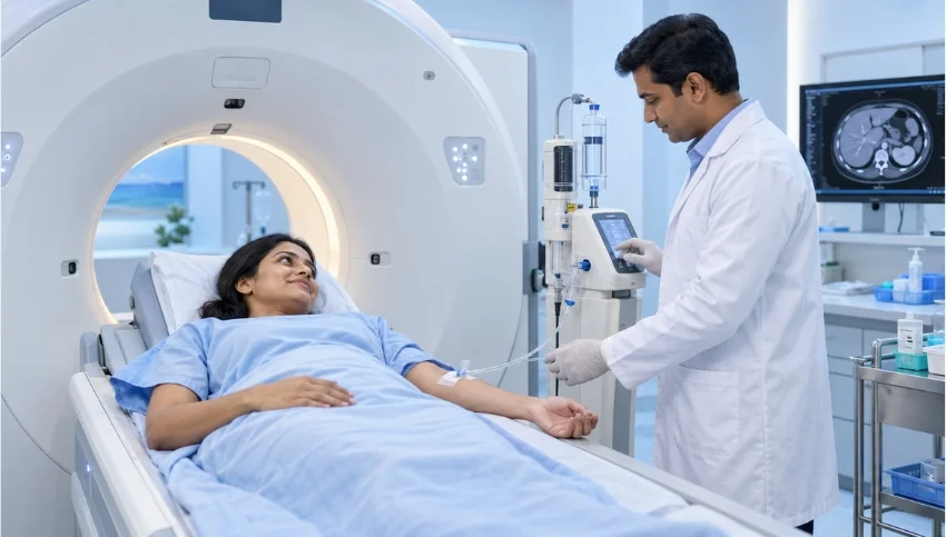
- Injected into a vein
- Introduced through the rectum
- Introduced into a specific anatomical space
- Administered directly into an area being evaluated during an image-guided examination
The radiology team will explain the procedure and administration method before the examination.
How Should You Prepare for a Contrast Procedure?
Preparation varies considerably depending on the specific examination.
Patients may receive instructions regarding:
- Fasting before the examination
- Food and fluid intake
- Regular medications
- Previous contrast reactions
- Kidney-related medical history
- Diabetes or other medical conditions
- Pregnancy or possible pregnancy
- Previous imaging reports
- Relevant laboratory tests when required
Patients should follow the specific preparation instructions provided by the imaging centre and referring doctor.
Do not assume that every Contrast Procedure requires fasting. Preparation requirements depend on the examination being performed.
Is Contrast Material Safe?
Contrast materials are generally considered safe when used appropriately, although mild or, rarely, serious reactions can occur.
Radiology departments follow established procedures to identify potential risks and respond to adverse reactions when they occur.
Before receiving contrast, patients should tell the radiology team about:
- Previous reactions to contrast material
- Known allergies
- Kidney disease
- Thyroid conditions
- Heart disease
- Diabetes
- Pregnancy or possible pregnancy
- Other significant medical conditions
- Previous relevant imaging examinations
This information helps the medical team determine the safest and most appropriate approach.
What Sensations Can Occur During Contrast Administration?
Some patients may notice temporary sensations depending on the contrast material and route of administration.
With certain iodine-based intravenous contrast examinations, a temporary warm or flushed feeling or metallic taste may occur.
With gadolinium-based MRI contrast, some patients may experience temporary coolness around the injection site.
These sensations are usually brief, but patients should immediately tell the imaging team if they experience unusual or concerning symptoms.
Contrast Procedures and Kidney Health
Kidney function can be an important consideration before selected contrast-enhanced examinations.
For some MRI examinations involving gadolinium-based contrast, the radiology team may ask about kidney disease and may require assessment of kidney function depending on the patient’s circumstances and the examination.
Patients should always disclose known kidney disease, kidney failure, kidney transplantation or other relevant medical conditions before contrast administration.
Contrast Procedures During Pregnancy
If you are pregnant or think you may be pregnant, inform your doctor and radiology team before the examination.
The decision to perform an imaging examination with contrast depends on the medical situation, the type of imaging required and the potential benefits and risks.
For MRI, gadolinium contrast is generally avoided during pregnancy unless the information is considered important enough to justify its use. For CT examinations, iodinated contrast may be used when clinically necessary after appropriate consideration by the treating team.
Never hesitate to tell the imaging team about a possible pregnancy before your examination.
Contrast Procedures vs Routine Imaging
Not every patient requires contrast-enhanced imaging.
Routine imaging without contrast may provide sufficient information for certain clinical questions, while contrast may be recommended when additional visualization is needed.
For example:
Without contrast:
The examination may provide adequate anatomical information for the clinical question.
With contrast:
The contrast material may improve the visibility or differentiation of selected organs, tissues or blood vessels.
The choice should be based on the patient’s clinical condition and the diagnostic question—not simply on whether contrast is considered more advanced.
What Should You Bring for a Contrast Examination?
Depending on the examination, it may be useful to carry:
- Doctor’s prescription or referral
- Previous imaging reports
- Previous scan images, if available
- Relevant medical records
- Medication information
- Kidney-function reports when requested
- Details of previous contrast reactions
Providing previous medical information can help the radiology team understand the clinical context of the examination.
Frequently Asked Questions
1. What are Contrast Procedures?
Contrast Procedures are imaging examinations in which a contrast material is used to improve the visibility of selected organs, tissues, blood vessels or internal structures.
2. Which contrast procedures are available at Super Scan?
Super Scan lists Barium Swallow, Barium Meal, Follow Through, Barium Enema, HSG, MCU, RGU, Sinogram and Fistulogram among its Contrast Procedures.
3. Is contrast required for every scan?
No. Contrast is not necessary for every imaging examination. Its use depends on the clinical question, examination type and information required.
4. Does contrast injection hurt?
An IV contrast examination involves placement of a small IV needle, which may cause brief discomfort. Some contrast materials can also cause temporary sensations such as warmth or a metallic taste.
5. Can I eat before a Contrast Procedure?
It depends on the specific examination. Some procedures have fasting or dietary instructions, while others may not. Follow the preparation instructions given for your particular examination.
6. Should I tell the radiologist about kidney disease?
Yes. Kidney disease can be relevant when certain contrast materials are being considered, particularly for selected MRI examinations using gadolinium.
7. Can Contrast Procedures be performed during pregnancy?
Some imaging examinations may be performed during pregnancy when medically necessary, but contrast use requires specific consideration. Always inform the doctor and imaging team about pregnancy or possible pregnancy before the examination.
8. Are Contrast Procedures safe?
Contrast materials are generally safe when appropriately selected and administered, although adverse reactions can occur. Serious reactions are uncommon, and imaging facilities maintain procedures for managing them.