Discover how advanced diagnostic imaging and radiology services such as MRI, CT, ultrasound, mammography, PET-CT and image-guided procedures support accurate medical evaluation.
Advanced Imaging. Experienced Expertise. Better Diagnostic Insights.
What Is an Advanced Diagnostic & Radiology Centre?
An Advanced Diagnostic & Radiology Centre is a specialized medical imaging facility where different technologies are used to help doctors evaluate diseases, injuries and other health conditions.
Modern diagnostic radiology can include several imaging methods, such as X-ray, CT, MRI, ultrasound, mammography and nuclear medicine. Each technology provides different information, and the appropriate examination depends on the patient’s symptoms and the clinical question being investigated.
An advanced imaging centre brings these technologies together with trained radiologists, technologists and supporting medical professionals to provide diagnostic information that can assist referring doctors with clinical decision-making.
Why Is Diagnostic Imaging Important?
Many medical conditions cannot be diagnosed accurately from symptoms and physical examination alone.
Diagnostic imaging can help doctors:
- Identify abnormalities inside the body
- Evaluate injuries and structural changes
- Assess organs and soft tissues
- Examine blood vessels and blood flow
- Support cancer evaluation
- Monitor certain diseases over time
- Guide selected minimally invasive procedures
- Provide information for treatment planning
The imaging method is selected according to the patient’s condition and the information the doctor needs.
Radiologists are physicians trained to interpret medical images from modalities including X-ray, CT, MRI, ultrasound, nuclear medicine and PET.
What Services Are Available at an Advanced Radiology Centre?
A comprehensive diagnostic imaging centre may offer multiple imaging technologies under one healthcare network.
At Super Scan Imaging Centres, Surat, the current service portfolio includes 3T MRI, CT Scan, Interventional Radiology, Ultrasound, 3D/4D Sonography, Color Doppler, Mammography, Digital X-Ray and nuclear imaging/PET-CT-related services.
The advantage of having multiple imaging options is that patients can be directed toward the examination most appropriate for their clinical requirement.
3T MRI for Detailed Soft-Tissue Imaging
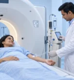
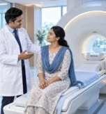
MRI uses a strong magnetic field and radiofrequency energy to create detailed images of internal body structures without ionizing radiation.
A 3T MRI system operates at a magnetic field strength of 3 Tesla and can provide detailed imaging for many clinical applications.
MRI may be used for evaluating areas such as:
- Brain and nervous system
- Spine
- Joints
- Muscles and ligaments
- Abdomen
- Pelvis
- Prostate
- Selected breast examinations
The exact MRI protocol depends on the body region and clinical indication.
Super Scan’s website identifies its 3T MRI service as part of its advanced diagnostic imaging offering.
CT Scan for Fast and Detailed Imaging
Computed tomography, or CT, uses X-rays and computer processing to produce cross-sectional images of the body.
One important advantage of CT is speed. Modern CT examinations can acquire detailed images rapidly, making CT useful in many emergency and non-emergency clinical situations.
CT may be used for:
- Trauma assessment
- Chest and lung imaging
- Abdominal evaluation
- Kidney stones
- Bone abnormalities
- Certain vascular examinations
- Cancer assessment and treatment planning
Super Scan currently highlights a 160 Slice CT system among its diagnostic imaging capabilities.
Because CT uses X-rays, radiation considerations are part of the examination decision. The radiation dose varies according to the type of CT examination and the body region being evaluated.
Ultrasound and Sonography for Real-Time Imaging
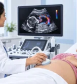
Ultrasound uses sound waves to create images of structures inside the body.
It can be used to examine a variety of areas, including:
- Abdomen
- Pelvis
- Liver
- Gallbladder
- Kidneys
- Thyroid
- Breast
- Prostate
- Soft tissues
- Pregnancy-related structures
Ultrasound does not use ionizing radiation and can provide real-time imaging. Super Scan’s ultrasound services include several organ-specific examinations as well as obstetric and paediatric ultrasound.
Color Doppler for Blood-Flow Assessment
Color Doppler is a specialized ultrasound technique that adds information about blood movement through vessels.
It can be used in selected examinations of:
- Arteries
- Veins
- Carotid vessels
- Abdominal vessels
- Selected organs
- Pregnancy-related blood-flow assessment
Doppler ultrasound can provide information that complements conventional grayscale ultrasound.
The appropriate Doppler examination depends on the blood vessel or organ being evaluated and the clinical question.
3D and 4D Sonography
3D and 4D sonography use advanced ultrasound techniques to create three-dimensional or real-time three-dimensional views in selected applications.
They are particularly associated with prenatal imaging, although the exact examination and clinical usefulness depend on the patient’s circumstances and the purpose of the scan.
Super Scan lists 3D/4D Sonography among its imaging services, including prenatal imaging applications.
Digital X-Ray for Routine and Specialized Imaging
Digital X-ray is one of the most widely used diagnostic imaging techniques.
It can help evaluate bones, the chest and other areas depending on the examination.
Digital imaging allows X-ray images to be captured and processed electronically, supporting efficient image access and reporting.
Although X-rays use ionizing radiation, the amount varies according to the examination. The decision to perform an X-ray should be based on the clinical need.
Mammography for Breast Imaging
Mammography uses specialized X-ray equipment to examine breast tissue.
It can be used for breast screening and diagnostic evaluation depending on the patient’s age, symptoms, risk factors and medical history.
Additional imaging such as breast ultrasound or MRI may be recommended in selected circumstances.
The appropriate breast imaging strategy should be determined by the treating doctor and imaging specialist.
PET-CT and Nuclear Medicine Imaging
Nuclear medicine provides information about physiological or biological processes by using radiotracers and specialized imaging equipment.
PET-CT combines PET imaging with CT to provide complementary functional and anatomical information.
Nuclear medicine can be used in the evaluation of selected cancers, heart conditions and other diseases.
Super Scan’s current website identifies Khatodara as the centre for PET-CT and Gamma services under Super Sahyog Nuclear Imaging.
The specific nuclear medicine examination depends on the disease being investigated and the information required by the treating team.
Interventional Radiology: Imaging With Minimally Invasive Procedures
Radiology is not limited to diagnostic imaging.
Interventional Radiology uses imaging guidance to perform selected minimally invasive procedures.
Depending on the clinical situation, image guidance may involve ultrasound, CT, X-ray or other imaging techniques.
Examples of image-guided procedures can include selected biopsies, fluid drainage and vascular procedures.
The purpose is to provide targeted treatment or obtain diagnostic samples while using imaging to guide the procedure accurately. RadiologyInfo describes interventional radiology as a field involving minimally invasive techniques performed with imaging guidance.
Super Scan’s current website also identifies interventional radiology as part of its service portfolio.
What Role Does a Radiologist Play?
An advanced imaging machine alone does not provide a complete diagnosis.
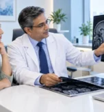
The radiologist plays an important role in selecting or planning appropriate imaging protocols, reviewing the images and interpreting the findings in the context of the patient’s clinical information.
Radiologists can also specialize in particular areas such as:
- Neuroradiology
- Musculoskeletal imaging
- Abdominal imaging
- Breast imaging
- Cardiovascular imaging
- Emergency radiology
- Interventional radiology
- Nuclear medicine
RadiologyInfo explains that radiologists are physicians trained to interpret multiple types of medical imaging and can work with referring doctors to determine appropriate imaging when needed.
Why Does Subspecialty Reporting Matter?
Different medical conditions can require different areas of radiological expertise.
For example, imaging of the brain, joints, abdomen, breast or cardiovascular system may involve different protocols and areas of specialization.
Super Scan states that its network includes experienced radiologists and subspecialty reporting, along with organ-specific expertise.
This approach can help ensure that imaging findings are interpreted in the appropriate clinical context.
What Is AI-Enabled Medical Imaging?
Artificial intelligence is increasingly being incorporated into medical imaging workflows.
AI-based tools may assist with aspects of image acquisition, reconstruction, workflow or analysis depending on the technology and clinical application.
However, AI should not be presented as a replacement for qualified medical professionals. The clinical interpretation of an examination remains dependent on the appropriate medical context, imaging quality and expert review.
Super Scan currently describes its network as including AI-enabled imaging systems.
Why Is Having Multiple Imaging Technologies Important?
Different imaging technologies answer different clinical questions.
For example:
MRI can provide detailed soft-tissue information.
CT can provide rapid cross-sectional imaging and is particularly valuable in many emergency settings.
Ultrasound can provide real-time imaging without ionizing radiation.
Mammography is specifically designed for breast imaging.
PET-CT can combine anatomical and metabolic information.
Digital X-ray can provide fast radiographic images for a variety of clinical indications.
There is therefore no single imaging technology that is universally best for every patient.
The appropriate examination depends on the clinical question, body region, symptoms, medical history and information required by the treating doctor.
What Should You Consider When Choosing a Diagnostic Imaging Centre?
Patients often search for an advanced diagnostic centre in Surat based only on the availability of a particular machine.
However, several factors can be relevant.
Range of Imaging Services
Having multiple modalities available can make it easier to access different examinations when clinically required.
Experienced Radiologists
Image interpretation is an important part of diagnostic imaging. Appropriate expertise can help connect imaging findings with the patient’s clinical information.
Imaging Technology
Modern imaging equipment can support high-quality examinations when used with appropriate protocols and trained personnel.
Patient Safety
Imaging examinations should be performed according to appropriate medical indications, safety protocols and examination-specific requirements.
Reporting and Digital Access
Efficient digital systems can help facilitate access to images and reports for referring doctors and patients.
Convenient Locations
Accessibility can be important when patients need imaging, follow-up examinations or multiple visits.
Advanced Diagnostic & Radiology Centre in Surat
Super Scan Imaging Centres has been providing diagnostic imaging services in Surat since 2007. The organization states that it was founded by Dr. Nipun Jindal and Dr. Ghanshyam Ardeshana and has continued to expand its imaging capabilities.
According to the current Super Scan website, the network has four imaging centres in Majura Gate, Lal Darwaja, Vesu and Khatodara.
Its current service portfolio includes advanced MRI, CT, ultrasound, 3D/4D sonography, Color Doppler, mammography, digital X-ray, interventional radiology and nuclear imaging/PET-CT services.
The network also states that its centres are connected through a digital imaging system for access to scans and reports.
When Should You Visit a Diagnostic Imaging Centre?
A doctor may recommend diagnostic imaging when symptoms, examination findings, laboratory results or medical history indicate that imaging could provide useful information.
Depending on the situation, imaging may be used for:
- Persistent or unexplained symptoms
- Suspected injury
- Neurological symptoms
- Abdominal or pelvic complaints
- Joint and musculoskeletal problems
- Cancer evaluation
- Follow-up of known conditions
- Screening when medically appropriate
- Pre-treatment or treatment-planning assessment
Patients should not select an imaging test solely based on an online search. The appropriate examination should be discussed with the treating doctor.
Can One Diagnostic Scan Detect Every Problem?
No.
Every imaging modality has strengths and limitations.
MRI, CT, ultrasound, X-ray, mammography and nuclear medicine provide different types of information. In some situations, more than one examination may be required.
For example, an ultrasound may identify an abnormality that later requires MRI or CT for further characterization.
Similarly, PET-CT may provide information that is different from conventional CT or MRI.
The purpose of using multiple technologies is not necessarily to perform more tests—it is to obtain the information needed to answer the clinical question.
Frequently Asked Questions
1. What is an advanced diagnostic and radiology centre?
It is a medical imaging facility offering multiple diagnostic technologies and radiology expertise to help evaluate diseases, injuries and other medical conditions.
2. Which services are available at Super Scan?
Super Scan currently lists 3T MRI, CT Scan, Interventional Radiology, Ultrasound, 3D/4D Sonography, Color Doppler, Mammography, Digital X-Ray and nuclear imaging/PET-CT-related services among its offerings.
3. Is MRI safer than CT?
MRI and CT use different technologies. MRI does not use ionizing radiation, while CT uses X-rays. However, “safer” cannot be judged independently of the clinical situation because the appropriate test depends on the medical question.
4. Which scan is best for diagnosis?
There is no single best scan for every condition. The appropriate imaging examination depends on the body part, symptoms, suspected condition and information required by the doctor.
5. Why do I sometimes need more than one imaging test?
Different imaging technologies provide different information. A second examination may be recommended to characterize a finding or answer a clinical question that the first examination could not fully address.
6. Does every diagnostic scan use radiation?
No. MRI and ultrasound do not use ionizing radiation. CT and conventional X-ray use X-rays, while nuclear medicine examinations use radiotracers. Radiation exposure varies according to the examination.
7. Does a radiologist interpret my scan?
A radiologist is a physician trained to interpret medical images and prepare imaging reports. Depending on the examination, nuclear medicine specialists and other appropriately trained professionals may also be involved.
8. Are advanced imaging tests always better?
Not necessarily. Advanced technology is useful when it provides information relevant to the clinical question. The most appropriate examination is determined by the patient’s medical circumstances.